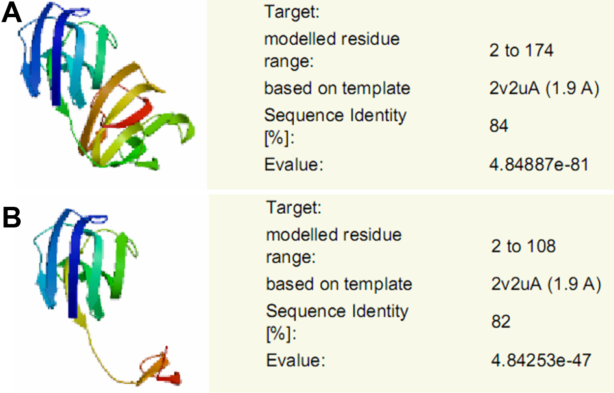
Figure 5. Structural modeling of the wild
type and mutant γC-crystallins. The structure modeling is based on the
X-ray determined coordinates of mouse γC-crystallin chain A using
SWISS-MODEL.
A:
A structural model of the wild type γC-crystallin with 84% sequence
identity is demonstrated.
B: A structural alteration of the
mutant γC-crystallin with 82% sequence identity is shown. Highly
symmetric structure of γC-crystallin is disrupted when 66 amino acids
are truncated from the COOH-terminus of γC-crystallin as result of
c.327C>A mutation.