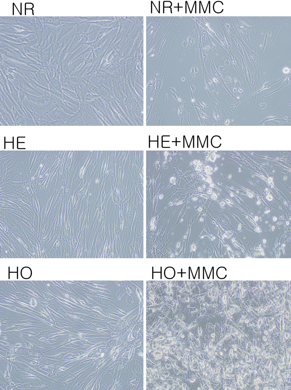
Figure 5. The effect of MMC on corneal
fibroblast apoptosis. Cells were incubated with 0.02% MMC for 6 h after
which they were stained with annexin V then avidin-horseradish
peroxidase (HRP) complex (1:300), and binding was visualized using a
0.05% diaminobenzidine/0.01% H2O2 solution. Cells
were photographed using a fluorescence microscope equipped with an
annexin V filter. Following MMC incubation, the GCD II homozygote cells
showed cytoplasmic shrinkage and perimembranous stippling prominently
compared with normal cells. MMC: mitomycin C, NR: normal, HE:
heterozygote, HO: homozygote.