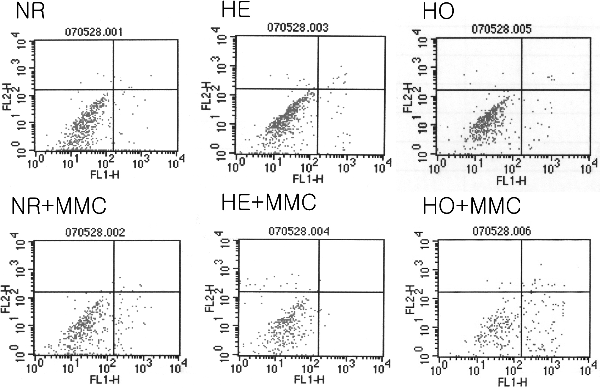
Figure 4. The effect of MMC on corneal
fibroblast apoptosis: FACS analysis. Cells were incubated with 0.02%
MMC for 6 h after which they were harvested, the DNA was stained with
propidium iodide, and the cells were analyzed using FACS. MMC increased
the proportion of apoptotic cells in GCD II homozygote cells. MMC:
mitomycin C, NR: normal, HE: heterozygote, HO: homozygote.