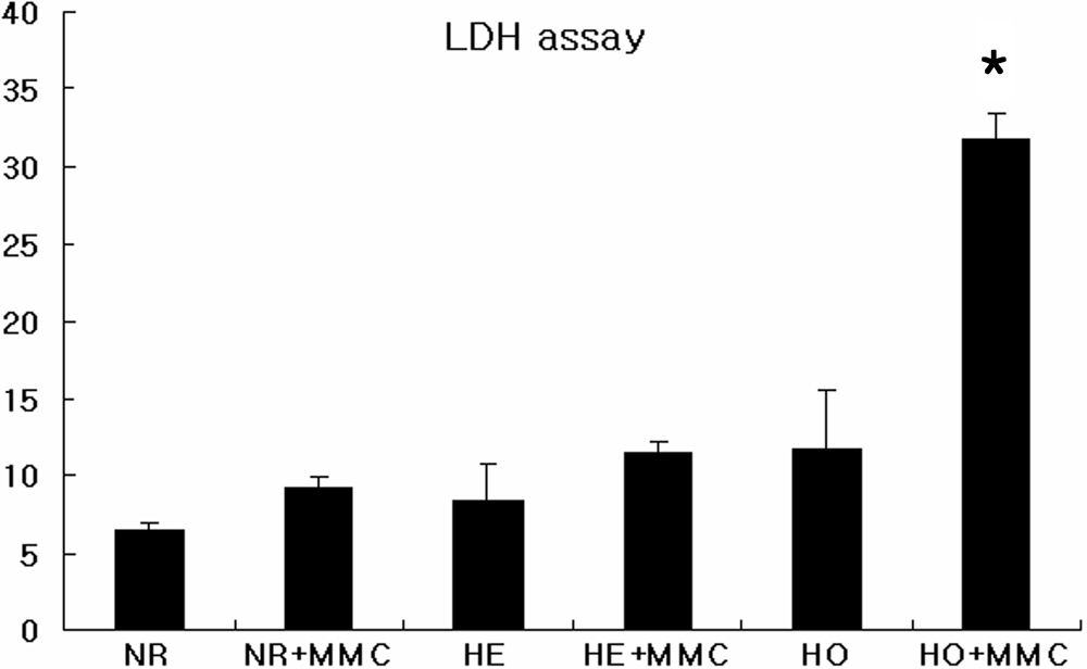
Figure 3. The effect of 0.02% MMC for 6 h
on corneal fibroblast viability. Cells were incubated with 0.02% MMC
for 6 h after which cell viability was measured using LDH assays. There
were fewer viable GCD II homozygote cells, compared to other cell
types. The asterisk indicates a p<0.05; ANOVA. MMC: mitomycin C, NR:
normal, HE: heterozygote, HO: homozygote.