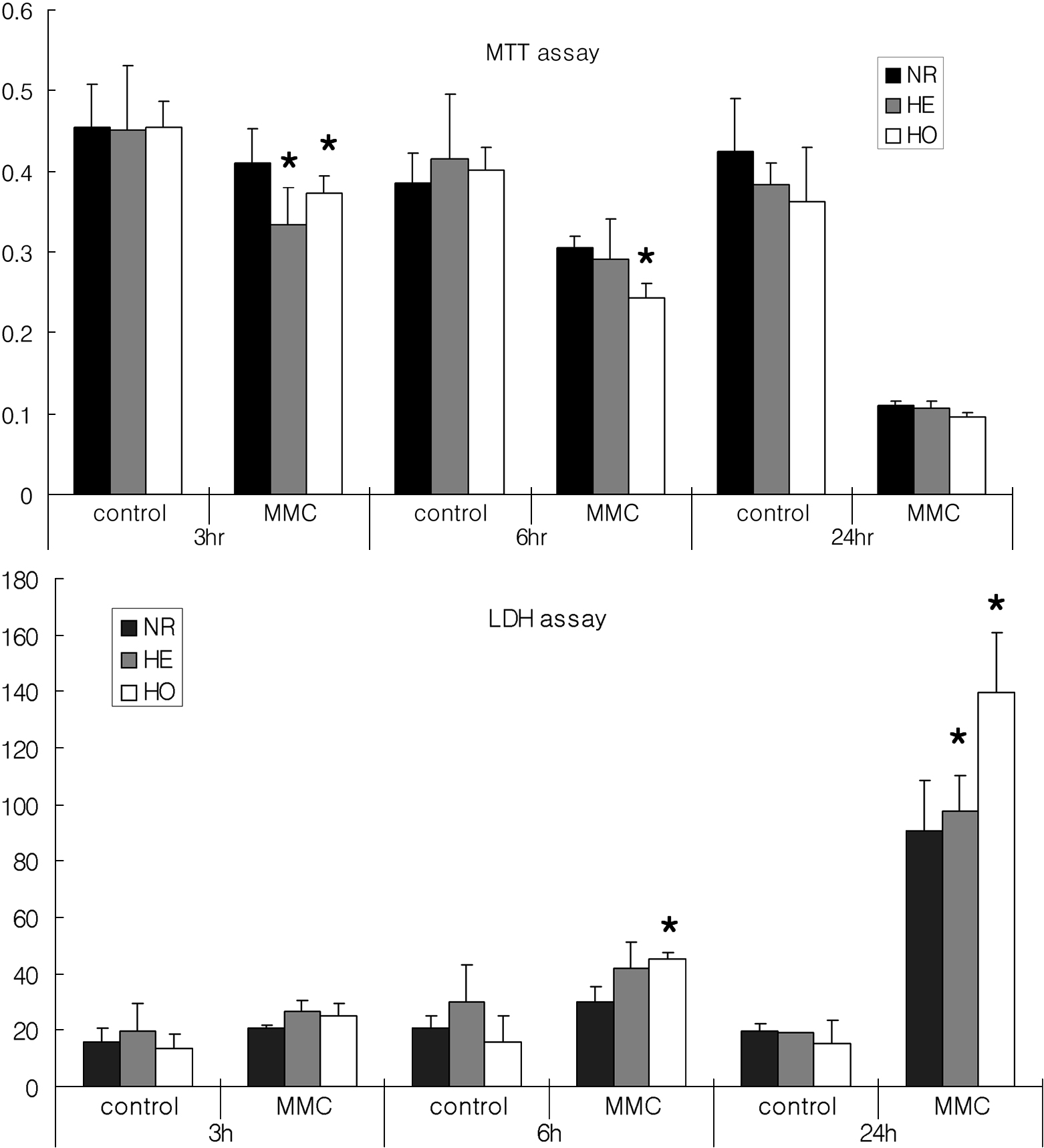
Figure 1. Time course for the effect of
0.02% MMC on corneal fibroblast viability. Corneal fibroblasts were
incubated with 0.02% MMC for various times after which cell viability
was measured using MTT and LDH assays. The MTT and LDH assays
demonstrated some minor differences, there were fewer viable GCD II
homozygote cells than normal cells. Upper panel=MTT assay, lower
panel=LDH assay. The asterisk indicates a p<0.05; ANOVA. MMC:
mitomycin C, control: without MMC, NR: normal, HE: heterozygote, HO:
homozygote.