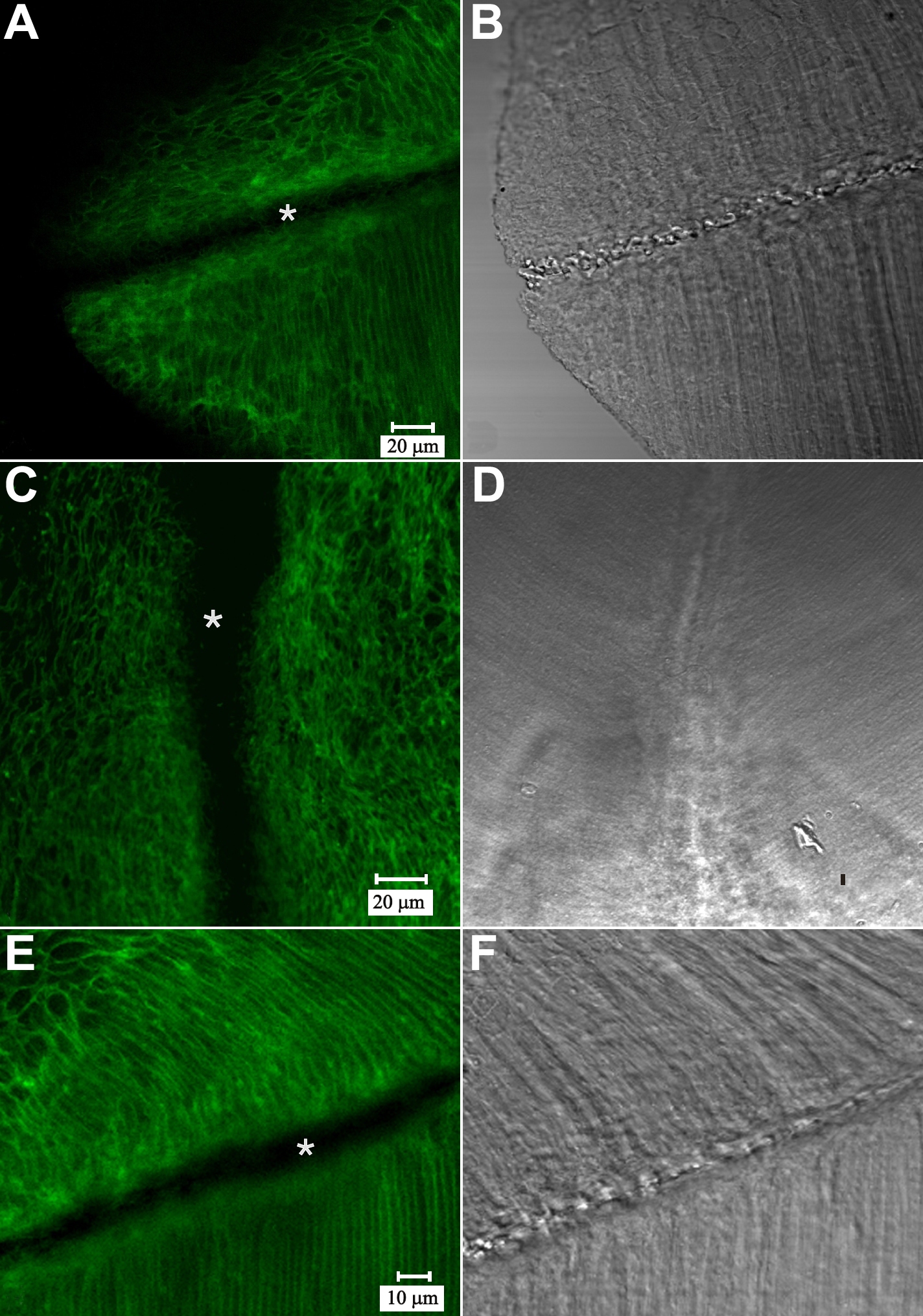
Figure 7. LSCM visualization of
pan-cadherin immuno-fluorescence at a posterior suture branch. Paired
fluorescent (A) and DIC (B) images of an oblique optical
section. A: Because the lens surface is curved, this optical
section demonstrates fluorescence due to cadherin family proteins in
both the BMC (upper portion) and in the lateral fiber membranes (lower
right). Notably, fluorescence is absent from the sutural region
(asterisk) A and B are at identical magnification. C-D:
Paired fluorescent (C) and DIC (D) LSCM images of a
posterior suture branch in a detergent-extracted section. Unmasking of
antigenic sites via this technique failed to reveal cadherin labeling
at, and adjacent to the posterior sutures (asterisk). C and D
are at identical magnification. E-F: Paired fluorescent (E)
and DIC (F) images of fully-elongated fibers that have detached
from the capsule and abutted to form a suture branch. Although the
posterior tips of these maturing fibers lack cadherin (asterisk),
strong cadherin fluorescence was apparent on the lateral fiber
membranes flanking the region where fiber ends abut and interdigitate. E
and F are at identical magnification.