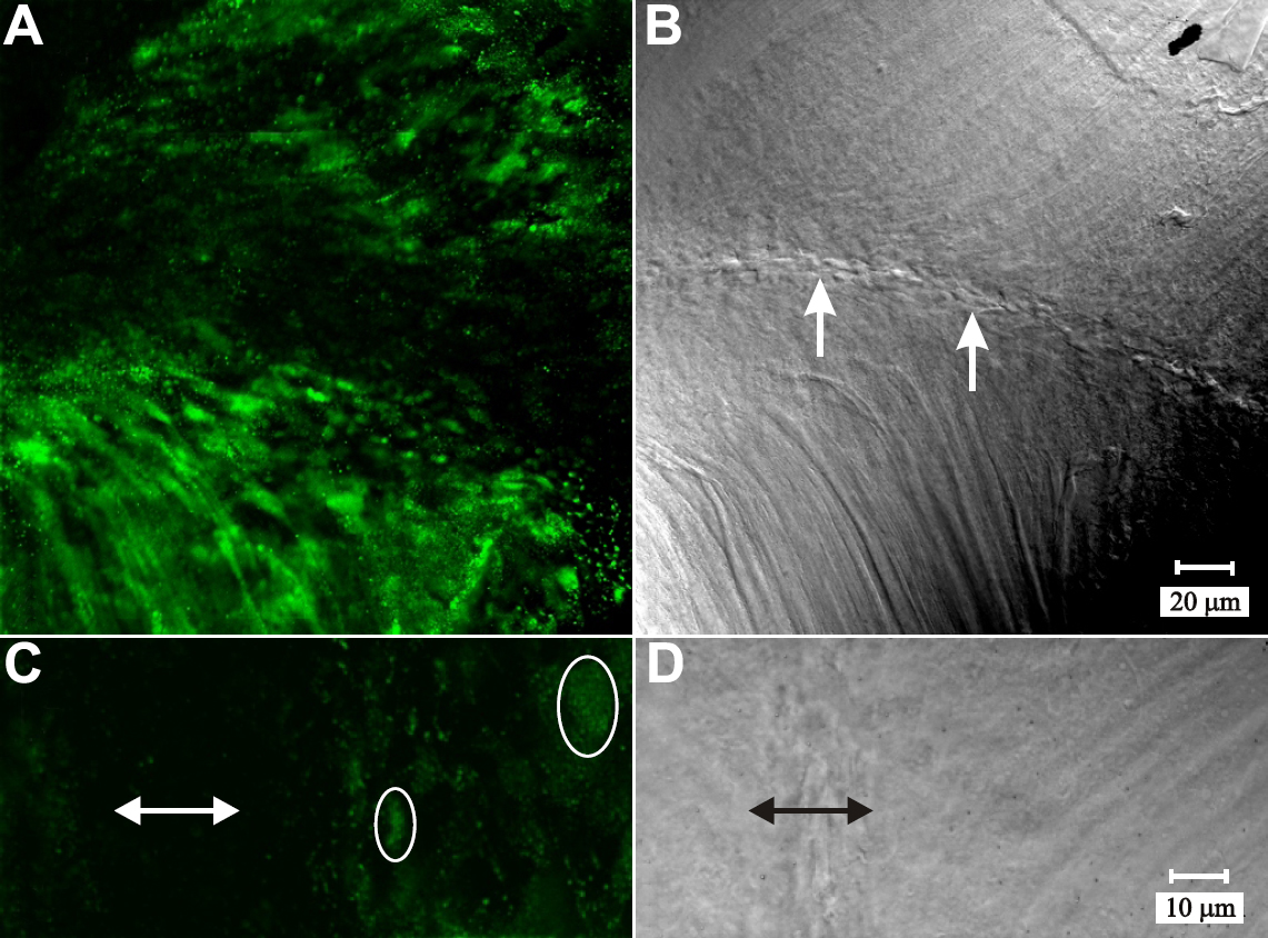
Figure 11. LSCM images of β1 integrin
immuno-fluorescence at posterior suture branches. A-B:
Paired fluorescent (A) and DIC (B) images of a posterior
suture branch (arrows). The marked reduction in β1 integrin
immuno-fluorescence was apparent in fiber ends at, and approaching the
sutural region of fiber end migration. A and B are at
identical maginificaiton. C-D: Higher magnification of
paired fluorescent (C) and DIC (D) micrographs at a
posterior suture branch. C: Approaching the suture, β-1
integrin was dispersed throughout the BMC (circled fiber end profiles).
D: Within the nascent sutures (double-headed arrow), the paucity
of label for β-1 integrin was apparent. C and D are at
identical magnification.