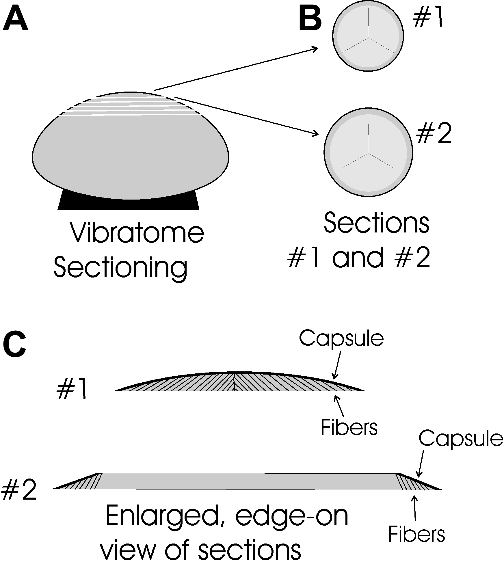
Figure 1. Diagrammatic representation of
the vibratome sectioning technique. A: Lenses were mounted on
the anterior surface and sectioned beginning at the posterior pole. B:
When viewed face-on, all sections contain portions of the suture
planes. Typically the first 1-3 sections contain elongating fiber ends
in the peri-sutural and sutural regions. C: An enlarged,
edge-on view of the first two sections demonstrates that migrating
fiber ends are present beneath the entire curved surface of the capsule
in section #1. However, in section #2 (and all subsequent sections)
elongating fiber ends are present only beneath the beveled edges of
sections covered by capsule.