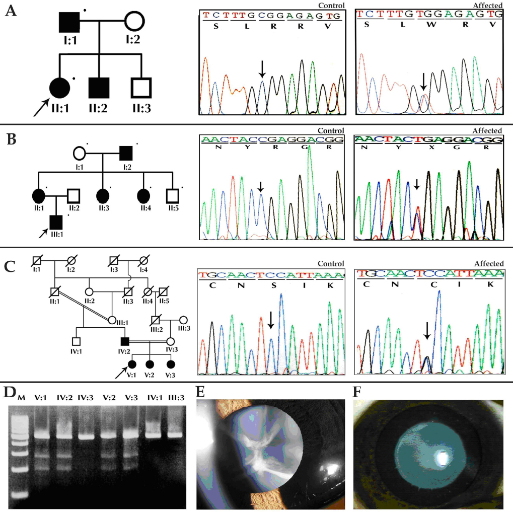
Figure 5. Mutation analysis of
γ-crystallin genes. A: Family CCW-33 shows an R168W mutation in
CRYGC. B: Family CCW-45 shows an R140X mutation in CRYGD.
C: Family CCW47 shows a S39C mutation in CRYGS. D:
RFLP analysis of CRYGS exon 1 shows the loss of HpyF10VI site
(mutant allele-274 bp and 204 bp, wild-type allele-478 bp). E:
Individual V:3 of family CCW47 shows sutural cataract. F:
Individual II:1 of family CCW33 shows lamellar cataract. M denotes 100
bp DNA ladder, and C denotes unrelated control.