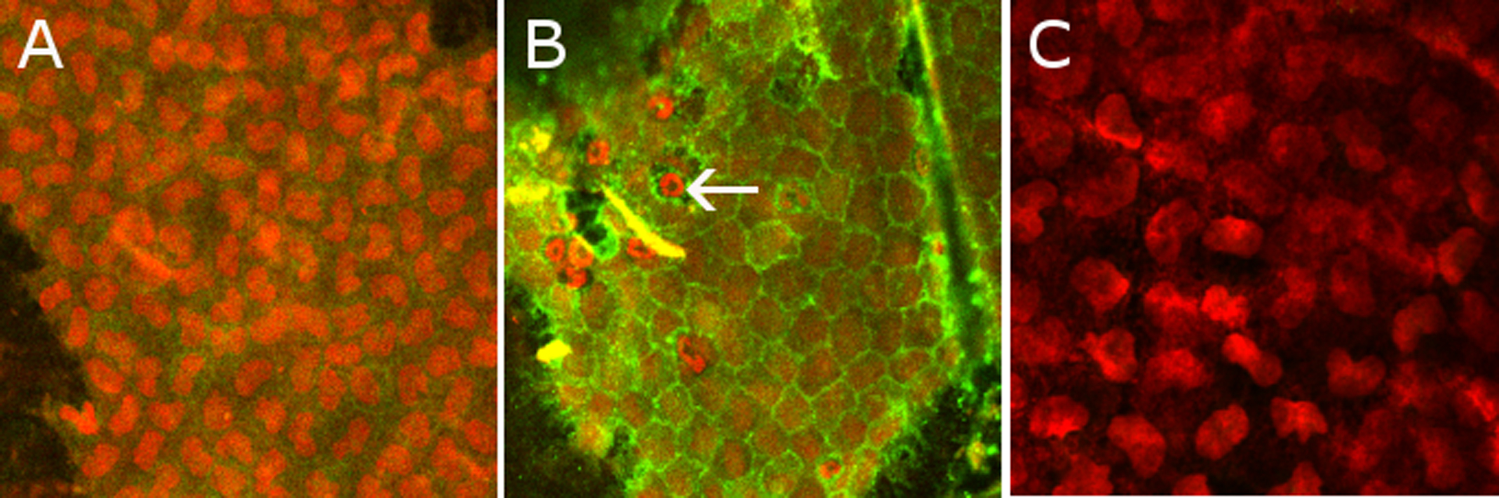
Figure 4. Double immunostaining with
anti-CD54 ICAM-1 (in green) and with propidium iodide (in red) on
corneas. The corneas were analyzed with a confocal microscope with a
200 X enlargement. Immunofluorescence of ICAM-1 is very weak in control
animals (A) and was increased 24 h after the LPS injection (B).
Some polymorphonuclear cells (arrow) are visible on the endothelium.
Double immunofluorescence with IgG1 mouse antibody for isotypic control
(revealed by secondary Alexa Fluor in green) and with propidium iodide
(in red) was performed in EIU (C) with a 250X enlargement.
Intensities of staining were the same for each assay.