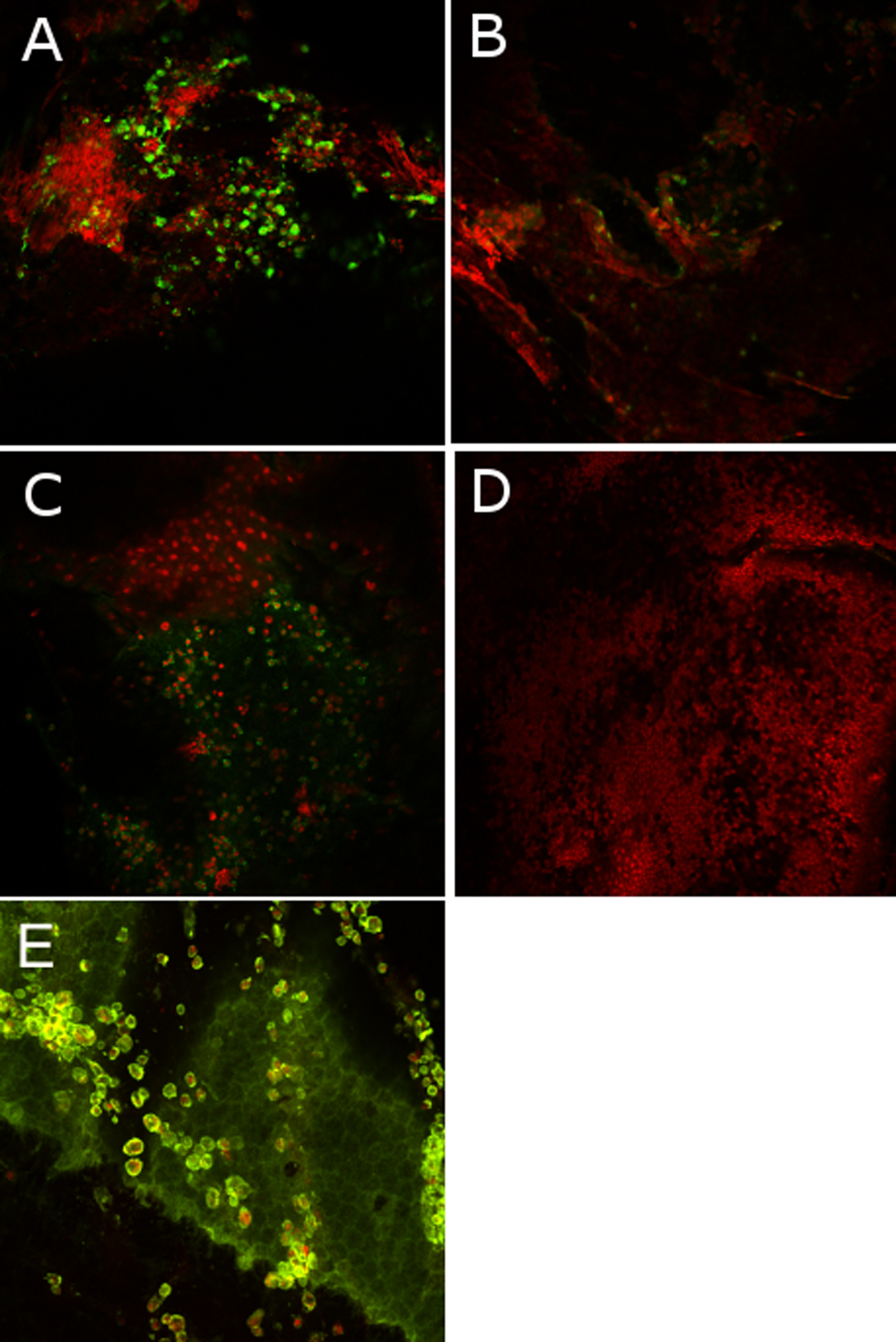
Figure 3. Immunofluorescence images on
flat-mounted corneas in a model of endotoxin-induced uveitis analyzed
with a 3D laser confocal microscope with a 20X enlargement.
Immunostainings with anti-CD68 for macrophages (A), anti-TCR
alpha/beta for lymphocytes (B), anti-MCA967 for granulocytes (C),
and IgG1 mouse antibody for isotypic control (D) were revealed
with secondary antibody, Alexa Fluor (in green) while the nuclear
chromatin was stained with propidium iodide (in red). A double
immunostaining with Alexa Fluor 488 phalloidin (in green), and antibody
to CD68 (in red) was performed to localize the macrophages among the
corneal layers, notably on the endothelium (E).