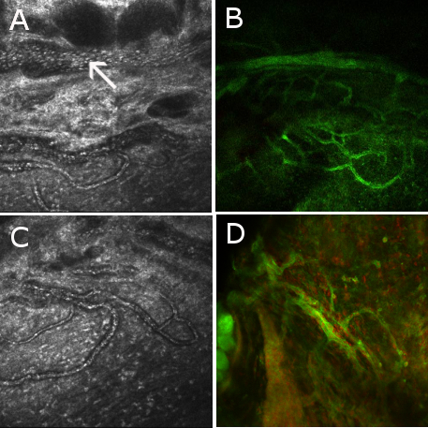
Figure 2. Correlation between in vivo
confocal microscopy and immunohistochemistry on peripheral
vascularisation in the corneal anterior stroma in a model of Endotoxin
Induced Uveitis. A: In vivo confocal microscopic images (400 μm
x 400 μm) of a circular peripheral network of vessels in the corneal
anterior stroma, deriving from a large circumferential vascular
structure resembling the major arterial circle of iris (arrow), are
shown of rats injected with LPS. Margination and diapedesis of
inflammatory cells can be visualized from these vessels toward the
anterior stroma (C). These peripheral corneal vessels can also
be shown in ex vivo immunostaining with phalloidin in the limbus of
flat-mount corneas from rats injected with LPS (B and D).