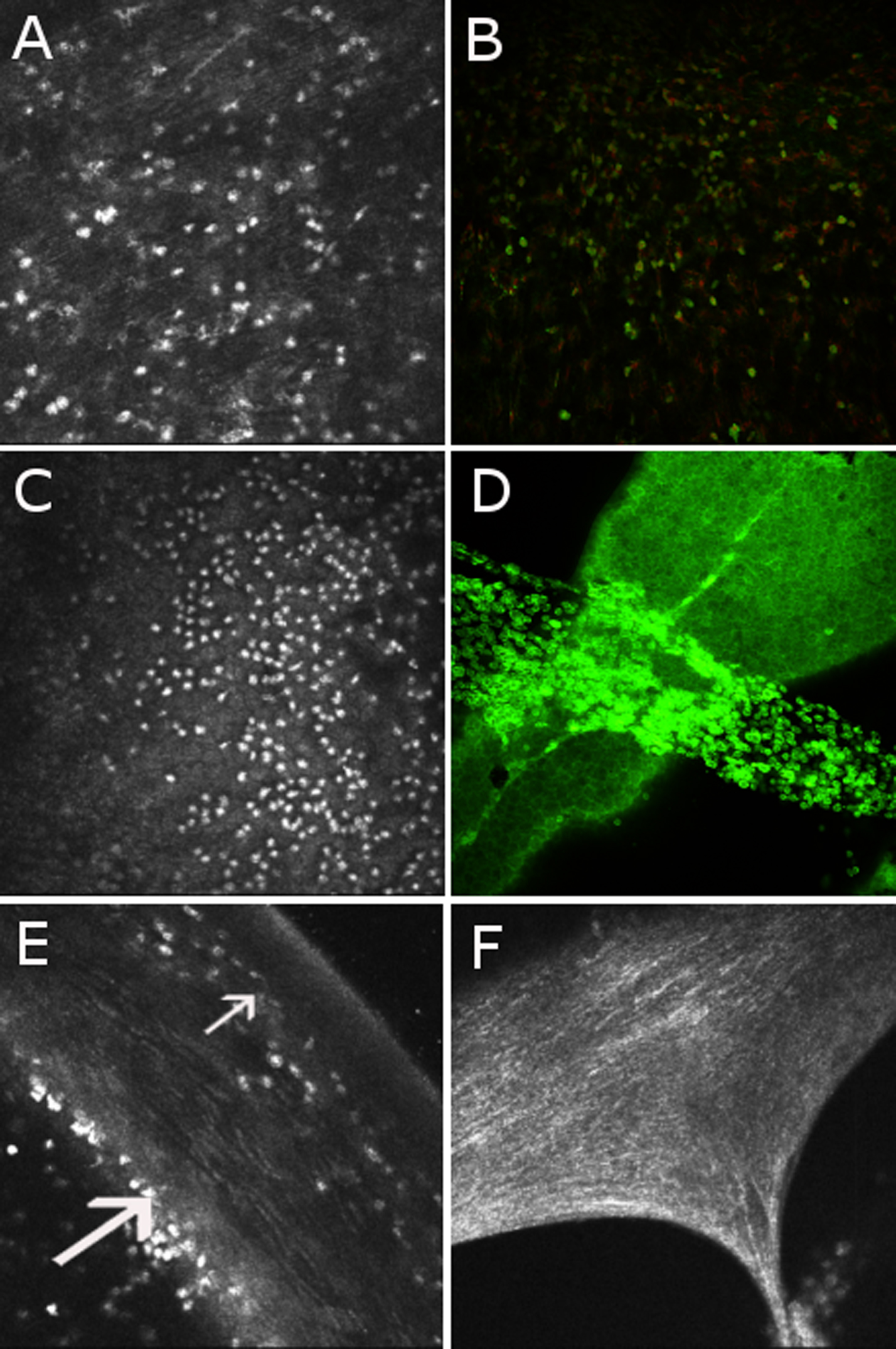
Figure 1. Correlation between in vivo
confocal microscopy with immunohistochemistry on cornea in a model of
Endotoxin Induced Uveitis. Heidelberg Retina Tomograph II (HRT II) in
vivo confocal microscopy images of the cornea (400x400 μm) in a model
of endotoxin-induced uveitis (A, C, E, and F)
are correlated with ex vivo immunostaining images with Alexa Fluor 488
phalloidin staining cytoskeleton (in green) and counterstained with
propidium iodide for nuclei (in red) on flat-mounted corneas that were
analyzed by confocal microscopy of the same animals with a 20X
enlargement (B and D). Inflammatory cells infiltrated
the anterior stroma (A and B) and accumulated on the
corneal endothelium, which appeared as a honeycomb-like mosaic (C
and D). The anterior stroma (small arrow) and endothelium
(large arrow) are shown on a corneal section by in vivo confocal
microscopy (E). A clot of fibrin was individualized in the
anterior chamber by HRT II (F).