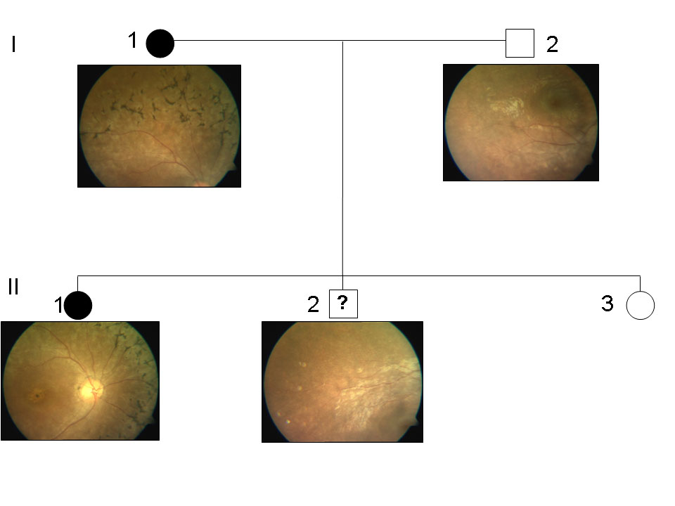
Figure 5. Fundus images of I-1, I-2, II-1,
and II-2 of family A. The fundus pictures from unaffected members I-2
and II-2 were normal. However, typical features of retinitis pigmentosa
could be well appreciated in affected members I-1 and II-1.The other
details of the members of family A are as follows; Individual I-1 was a
24-year-old female with a history of night blindness since 8 years. Her
visual acuity was counting fingers at 4 meters, and was not improving
with glasses (NIG). Fundus examination revealed arteriolar attenuation,
bony spicules with degenerative macular changes, and disc pallor. The
electroretinogram (ERG) was nonrecordable, fields were grossly
defective. Individual, I-2 was a 32-year-old male with a vision of 6/6
and normal fundus. Individual II-1 (proband) was a 5-year-old female
with a complaint of night blindness since 6 months. Fundus revealed
attenuated vessels, normal disc, dull foveal reflex, and altered
retinal sheen. The ERG was nonrecordable in both the eyes. Individuals
II-2 was a 3.5-year-old male with normal vision and fundus. Individual
II-3 is a 2-year-old female with normal fundus. The ERG and visual
field test could not be done due to pediatric age.