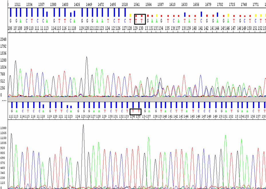
Figure 2. Electropherograms displaying a
novel PRPF31 mutation, p.Lys120GlufsX122. Genomic DNA sequences
(reverse primer) of a part of PRPF31 from a patient with the
p.Lys120GlufsX122 mutation (top) and from a normal subject (bottom).
The rectangular box shows the position of a heterozygous deletion of
two nucleotides at codon 120 (c. c.358_359 del AA, but the sequence
shows the reverse sequence boxed as TT).