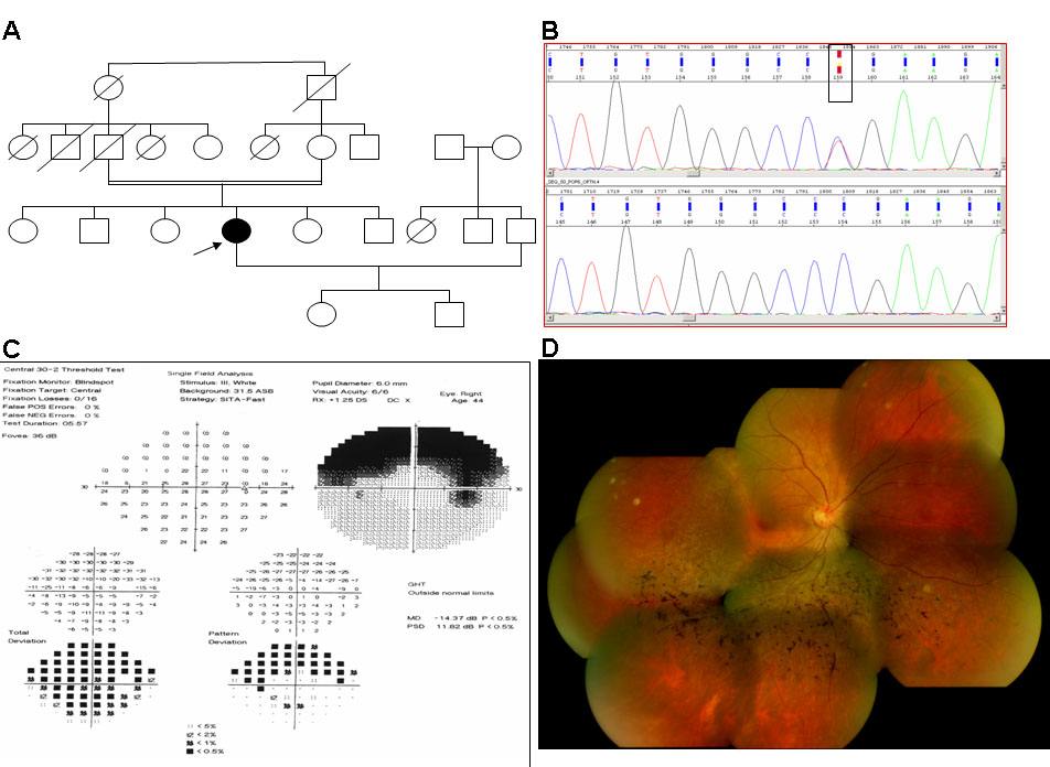
Figure 1. Clinical and molecular features
of the proband (L2:50) with the p.Gly106Arg mutation. A:
Pedigree showing the isolated form of the disease. B: Genomic
DNA sequences (reverse) of a part of the RHO gene of L2:50
displaying the p.Gly106Arg mutation (top) and of a normal subject
(bottom). The rectangular box shows the position of a heterozygous
change at nucleotide 316 (c.316G/A, but the sequence shows the reverse
sequence boxed as Y). C: Visual field test reveals a sectoral
form of RP. D: Fundus (right eye) photograph showing mild
retinitis pigmentosa changes.