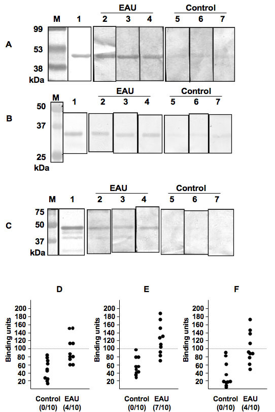
Figure 2. The presence of antibodies to
bAct, EsteD, and BB-CK in EAU mice was confirmed by 1D-WB and ELISA. A-C:
Protein (2 μg) was subjected to 12.5% SDS–PAGE, transferred onto
nitrocellulose membranes, then incubated with 1:200 sera from
experimental autoimmune uveoretinitis (EAU) or control mice and 1:2000
horse radish peroxide (HRP) antimouse IgG as described in Methods. EAU
serum samples were 8/10 positive in bAct (A), 6/10 positive in
EsteD (B), and 4/10 positive in brain-type creatine kinase
(BB-CK; C). D-F: Antibody titer of the sera from the
EAU or control mice was determined by ELISA. The antibody titer was
calculated as binding units according to the formula shown in Methods.
EAU serum samples were 4/10 positive in bAct (D), 7/10 positive
in EsteD (E), and 4/10 positive in BB-CK (F). Lane 1 is
ponseau S staining, lane 2-4 are membranes incubated with EAU sera, and
lane 5-7 are membranes incubated with control sera. In the figure, M
represents molecular weight marker.