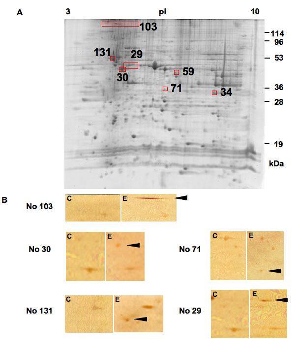
Figure 1. Detection of autoantigens in
experimental autoimmune uveoretinitis (EAU).
A: Shown is a
two-dimensional gel image of murine retinal proteins stained with SYPRO
Ruby. Approximately 2,000 spots were observed. Among the SYPRO Ruby
stained protein spots, the 2D-WB positive spots either in control or
EAU were randomly numbered. The numbers and the positions of the seven
candidate autoantigens in EAU on a SYPRO Ruby stained gel are shown in
panel
A. The spot numbers of the candidate autoantigens are
common between panel
B,
Table 1, and
Table 2.
B: Extracted murine retinal proteins were separated by
two-dimensional electrophoresis, transferred onto nitrocellulose
membranes. Western blotting was performed using sera from EAU or
control mice. Representative membranes reacted with sera from complete
Freund's adjuvant-treated control mice are shown in subpanel
C,
and those reacted with sera from EAU mice are shown in subpanel
E.
Each set of
C and
E subpanels shows the corresponding
area. Arrowheads indicate the position of each candidate autoantigen on
the EAU membranes. membranes.