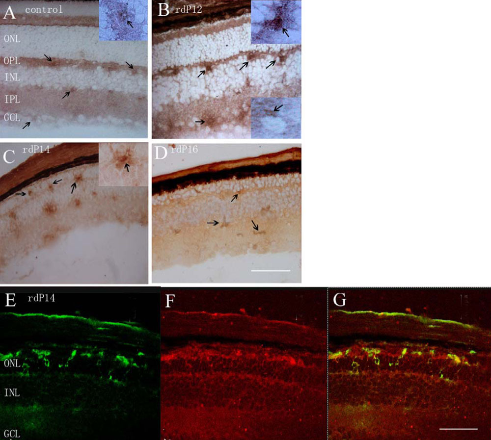
Figure 1. Immunochemical localization of
NF-κBP65 in rd retinas and controls. A: In control
retinas, weak immunoreactivity was present in the cytoplasm of cells in
the ganglion cell layer (GCL), inner plexiform layer (IPL), and outer
plexiform layer (OPL). The inserted picture showed magnified positive
cells in the OPL. B: In the rd retina at P12, P65
immunoreactivity peaked, and prominent nuclear labeling of the cells
was evident in the OPL. P65 immunoreactivity remained in the cytoplasm
in the cells of GCL. The inserted picture in the upper right angle
showed a magnified cell with nuclear labeling in the OPL. The inserted
picture in the lower right angle showed a magnified cell with cytoplasm
labeling in the GCL C: In the rd retina at P14,
migration of the cells with intense nuclear labeling was seen toward
the outer nuclear layer (ONL). The inserted picture showed a magnified
cell with nuclear labeling in the ONL. D: In the rd
retina at P16, P65 immunoreactivity was still prominent, but the cells
with nuclear labeling were hardly seen. E-G: Double labeling of
NF-κB P65 and CD 11b in the rd retina at P14 showed
co-localization of NF-κB in microglial cells in the outer retina. E:
Microglial cells were shown in green color; F: NF-κB P65
immunoreactivity was shown in red color. G: Expression of NF-κB
P65 in the microglial cells was shown in orange color. The arrow shows
positive labeling. In the figure, the inner nuclear layer is
abbreviated INL. Scale bar equals 100 μm.