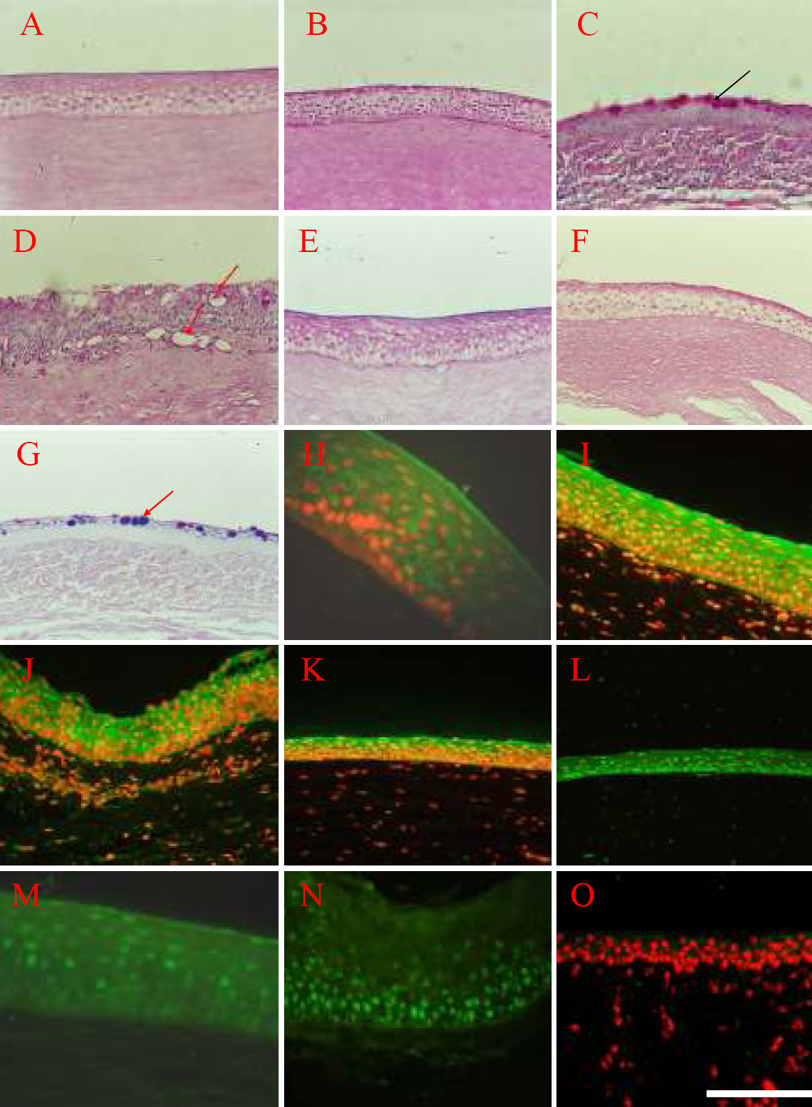
Figure 5. Epidermal adult stem cells can
be converted into corneal epithelium by corneal stroma. A: PAS
staining of the normal cornea epithelium showed no goblet cells, 200X. B:
PAS staining of the reconstructed cornea epithelium also show no goblet
cells, 200X. C-D: PAS staining shows the invaded goblet cells
in the corneal surface of control group, 200X. The black arrow in C
is pointing to the invaded goblet cells, the red arrow in D is
pointing to the blood vessels. E: Normal cornea is shown, and
the AB-PAS staining is red, characteristic of neutral mucus, 200X. F:
The reconstructed cornea is shown, and the AB-PAS staining is also red,
characteristic of neutral mucus, 200X. G: AB-PAS staining show
goblet cells as purple, which is characteristic of mixed mucus, 200X.
The red arrow in G is pointing to the goblet cells in the
corneal epithelium of control group which were stained purple by the
AB-PAS staining method. The results of immunohistochemistry showed that
normal cornea epithelium (H) expressed CK3 (I.BB.787), 400X, as
well as reconstructed cornea epithelium (I) expressed CK3, 400X.
Both normal cornea epithelium (J) and reconstructed cornea
epithelium (K) expressed CK12 (SC-17099) which is a specific
marker for differentiated corneal epithelium, 400X. Furthermore, both
of normal cornea epithelium (L) and reconstructed cornea
epithelium (M) expressed PAX-6 (AB5409), 400X. Although the
reconstructed epithelium of control group (N) expressed PAX-6
(AB5409), it did not express CK3 (O), showing that this kind of
epithelium derived from the control group did not share the character
with normal corneal epithelium, 200X. The scale bar represents 100 μm.