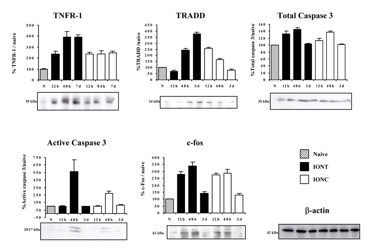
Figure 3. Time course regulation of cell
death–related proteins in naïve and optic nerve injured retinas.
Western blot time course analyses showing the regulation of tumor
necrosis factor receptor superfamily member 1a (TNFR1a), tumor necrosis
factor receptor type 1, associated death domain (TRADD), total Caspase
3, active Caspase 3, and c-fos in naïve, intraorbital nerve transection
(IONT)-, and intraorbital nerve crush (IONC)-injured retinas. Graphs
show quantification of protein signals (n=4 animals per lesion and time
point, western blots were replicated three times). The signal from
injured retinas is referred to the naïve signal, which was arbitrarily
considered 100%. To verify the amount of loaded protein, western blots
were incubated with β-actin (an example is shown). Error bars show the
standard error of the mean (SEM) for each experiment.