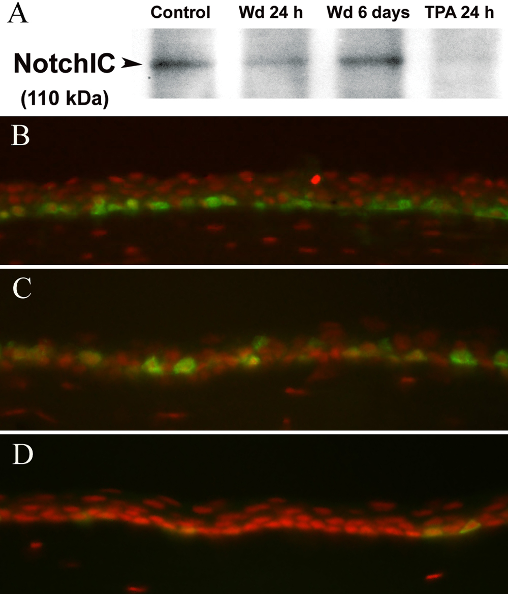
Figure 8. Western blotting for the cleaved
Notch intracellular domain. A: The results demonstrate reduced
levels of Notch intracellular domain (NotchIC) at 24 h after wounding
(Wd) and after TPA treatment. Immunofluorescence staining for Ki67
(green) demonstrates the number of proliferating basal cells to be
highest in the TPA-treated cornea at 24 h (B) followed by the
wounded cornea at 24 h (C) and a normal unwounded cornea (D).
The nuclei are stained red with propidium iodide.