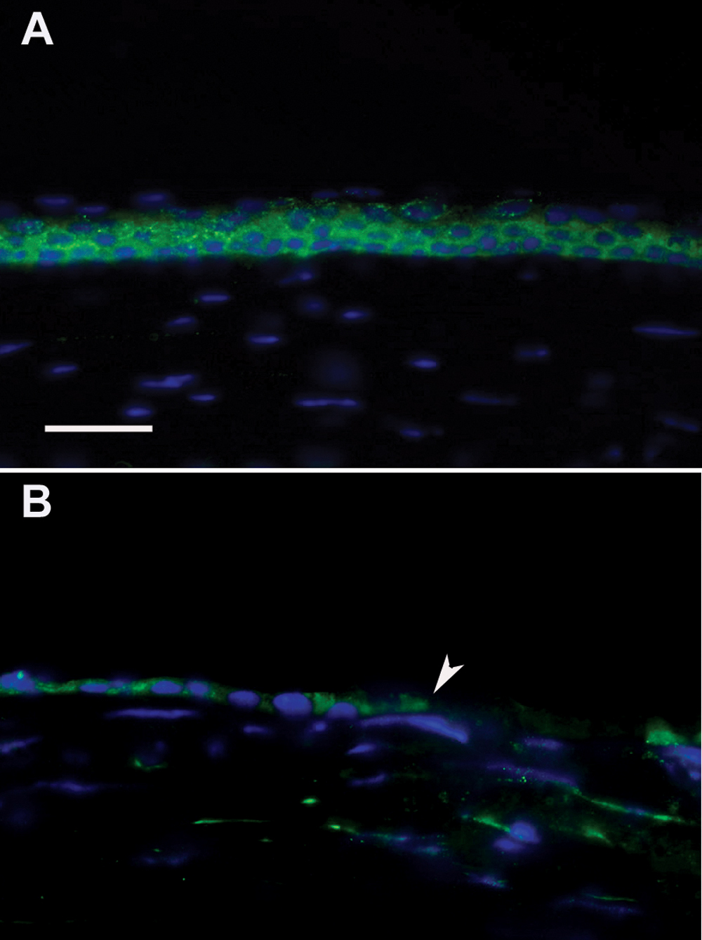
Figure 6. Immunofluorescence for Notch1 in
the cornea of a wounding mouse model. A: Notch1 expression is
noted predominantly in the basal and early suprabasal cells in
unwounded mouse corneas. B: Scattered expression of Notch1 is
seen in the new epithelium near the leading edge (arrowhead) 24 h after
wounding. Nuclei were stained with DAPI in blue. Bar=30 μm.