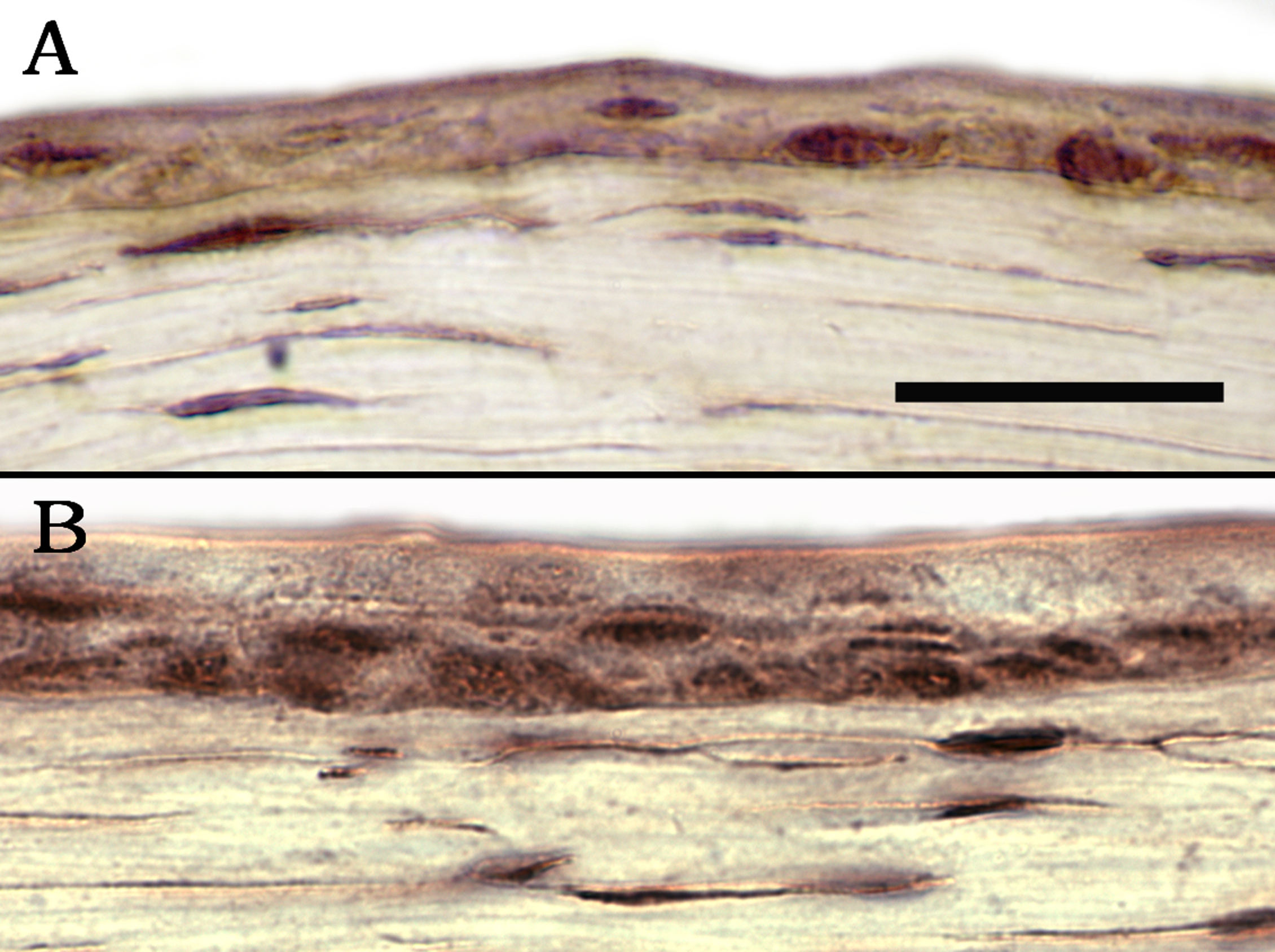
Figure 5. Immunohistochemical staining for
NotchIC in the cornea of a wounding mouse model. A: NotchIC is
seen in just a few basal and some suprabasal cells 24 h after wounding.
B: In the unwounded control mouse cornea, NotchIC staining is
demonstrated in the basal and suprabasal cells of the corneal
epithelium. The NotchIC staining is stronger in the unwounded cornea
(compare B versus A). Bar=30 μm.