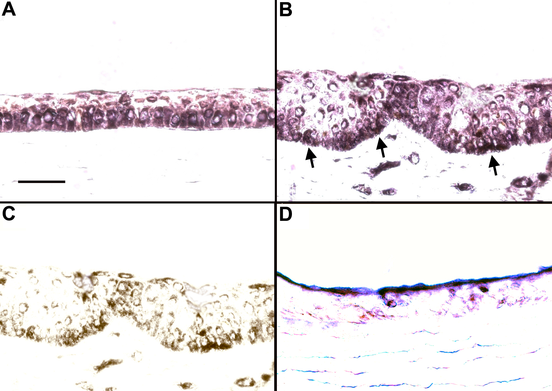
Figure 3. In situ hybridization of the
human Hes 1 gene. In both the central corneal epithelium (A)
and the limbal epithelium (B), expression of Hes1 is
seen predominantly in the basal layer with some extension into the
suprabasal layers. The intensity of the staining was slightly lower in
the limbus. The areas with darker staining observed in the limbal basal
epithelium (arrows) actually represent underlying pigmentation of the
tissue. The pigmented cells in the limbus are seen more clearly in C,
which represents the same section in B without the purple
staining. Minimal non-specific staining is observed in the sense
control in the central cornea (D). Bar=30 μm.