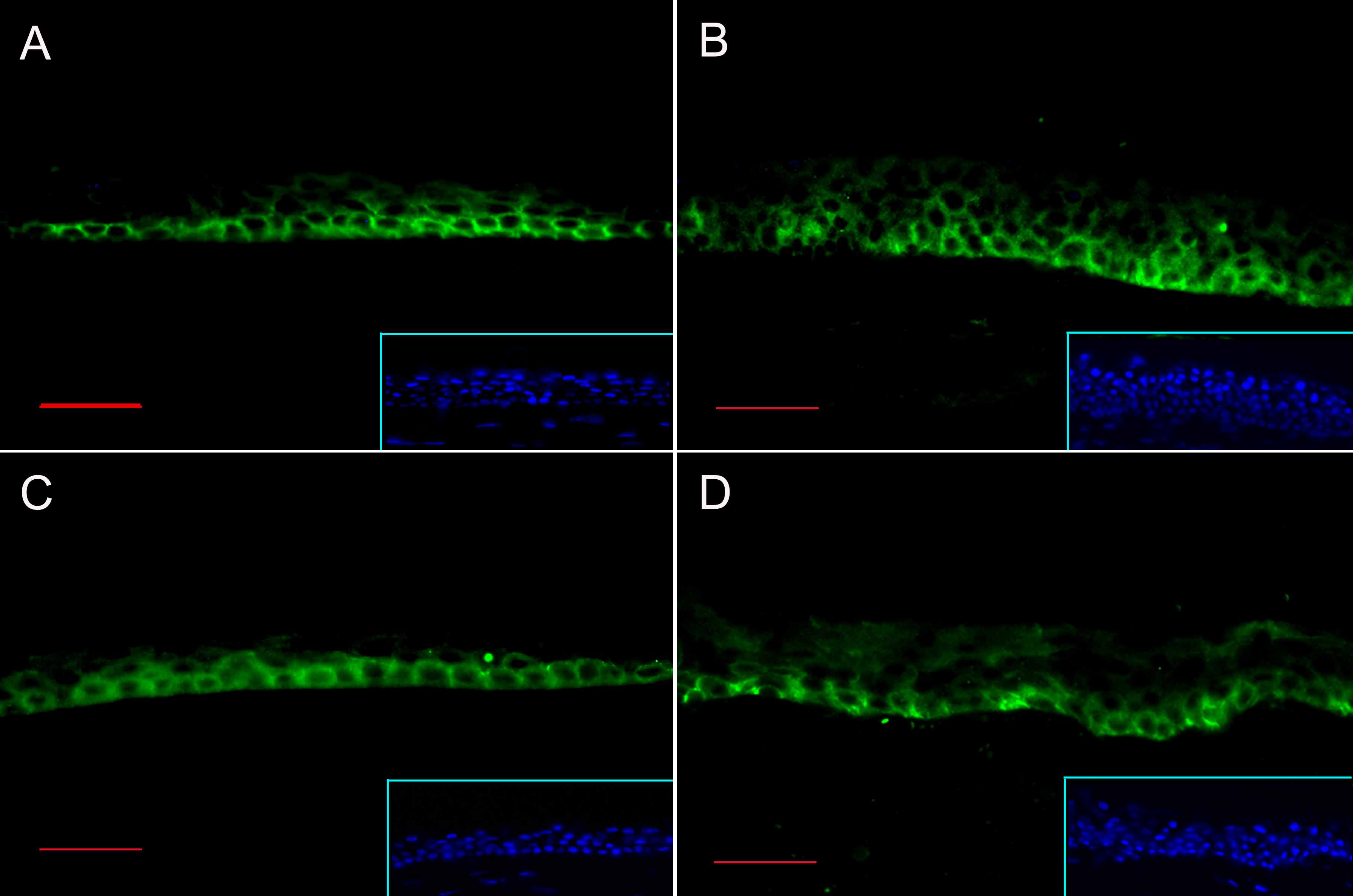
Figure 2. Immunofluorescence staining for
Notch1 and Jagged1. Notch1 (A and B) and Jagged1 (C
and D) staining is noted in the basal and immediate suprabasal
layers of central human cornea (A and C) and limbus (B
and D). Inset show the nuclear staining of the sections with
DAPI. Bar=30 μm.