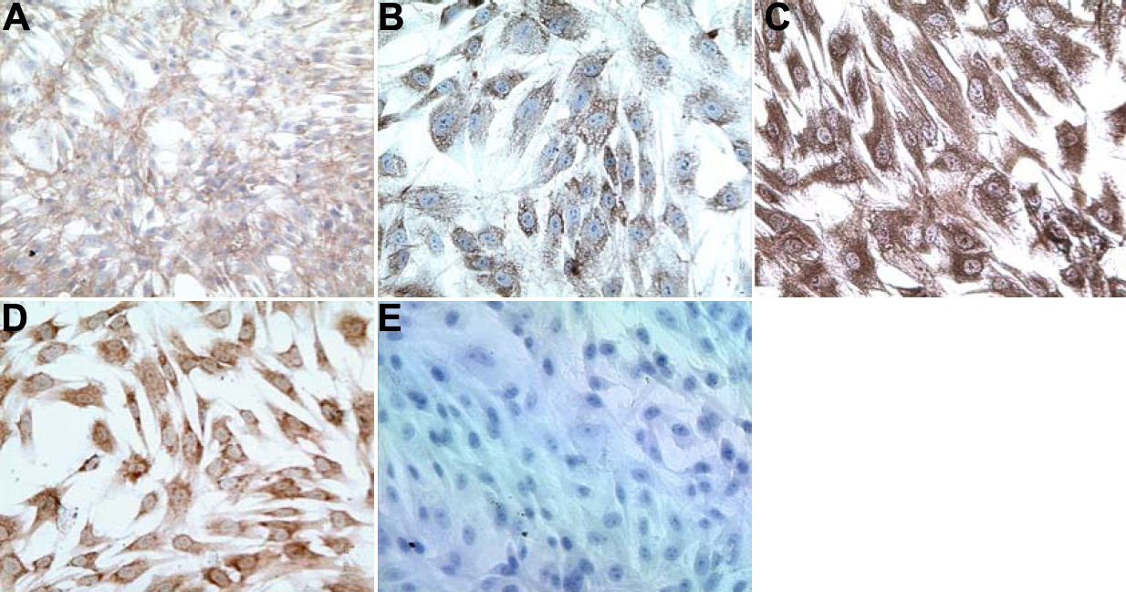
Figure 1. Immunohistochemical
characterization of primary trabecular meshwork cell cultures.
Immunohistochemical assays on the TM cells were positive for
fibronectin (A), laminin (B), vimentin (C),
neuronal specific enolase (D) and negative for the endothelial
cell marker, factor VIII (E), which indicated that the primary
cultures obtained from normal and POAG individuals were indeed TM cells.