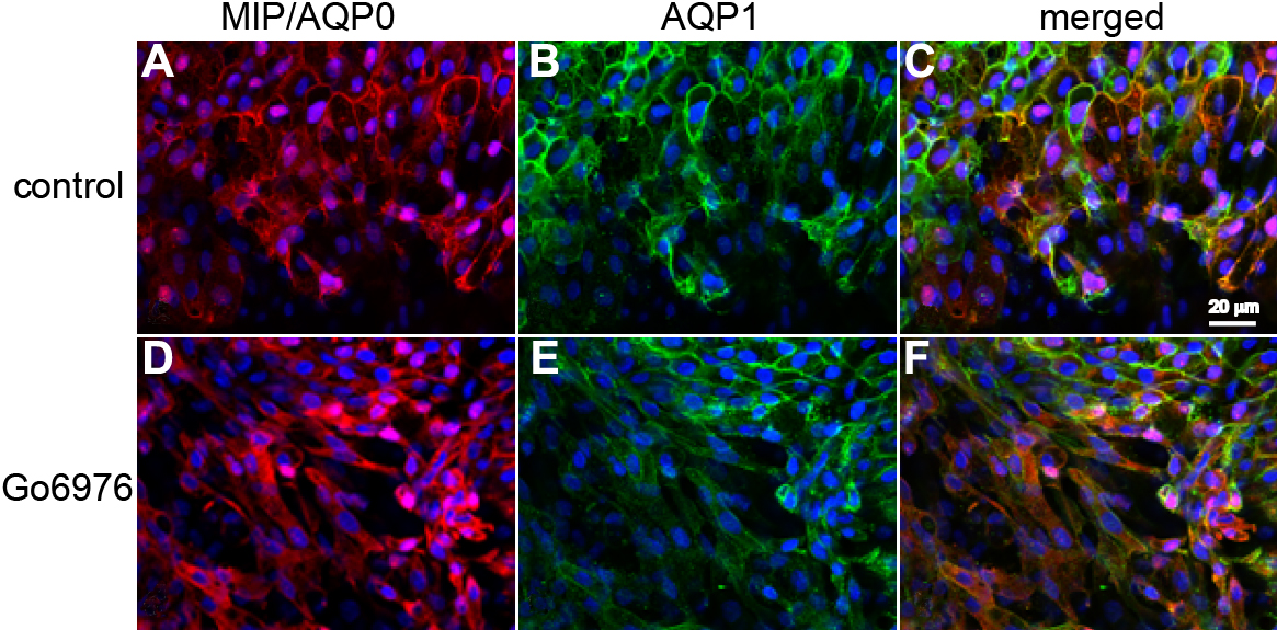
Figure 3. PKC inhibitor perturbs MIP/AQP0
membrane localization without affecting AQP1 membrane localization in
differentiating lens explants. Images are shown of MIP/AQP0
immunofluorescence (red; A) and AQP1 immunofluorescence (green;
B) in rat lens epithelial explants cultured in 100 ng/ml FGF-2
and 0.2% DMSO for 72 h (control, as described in Methods). Images are
shown of MIP/AQP0 immunofluorescence (red; D) and AQP1
immunofluorescence (green; E) in rat lens epithelial explants
cultured in 100 ng/ml FGF-2 in the presence of 4 μM Go6976 plus 0.2%
DMSO for 72 h (as described in Methods). C and F show
the merged images of MIP/AQP0 immunofluorescence and AQP1
immunofluorescence and the DAPI nuclear staining in controls (A
and B) and Go6976 experiments (D and E),
respectively. Scale bars represent 20 μm.