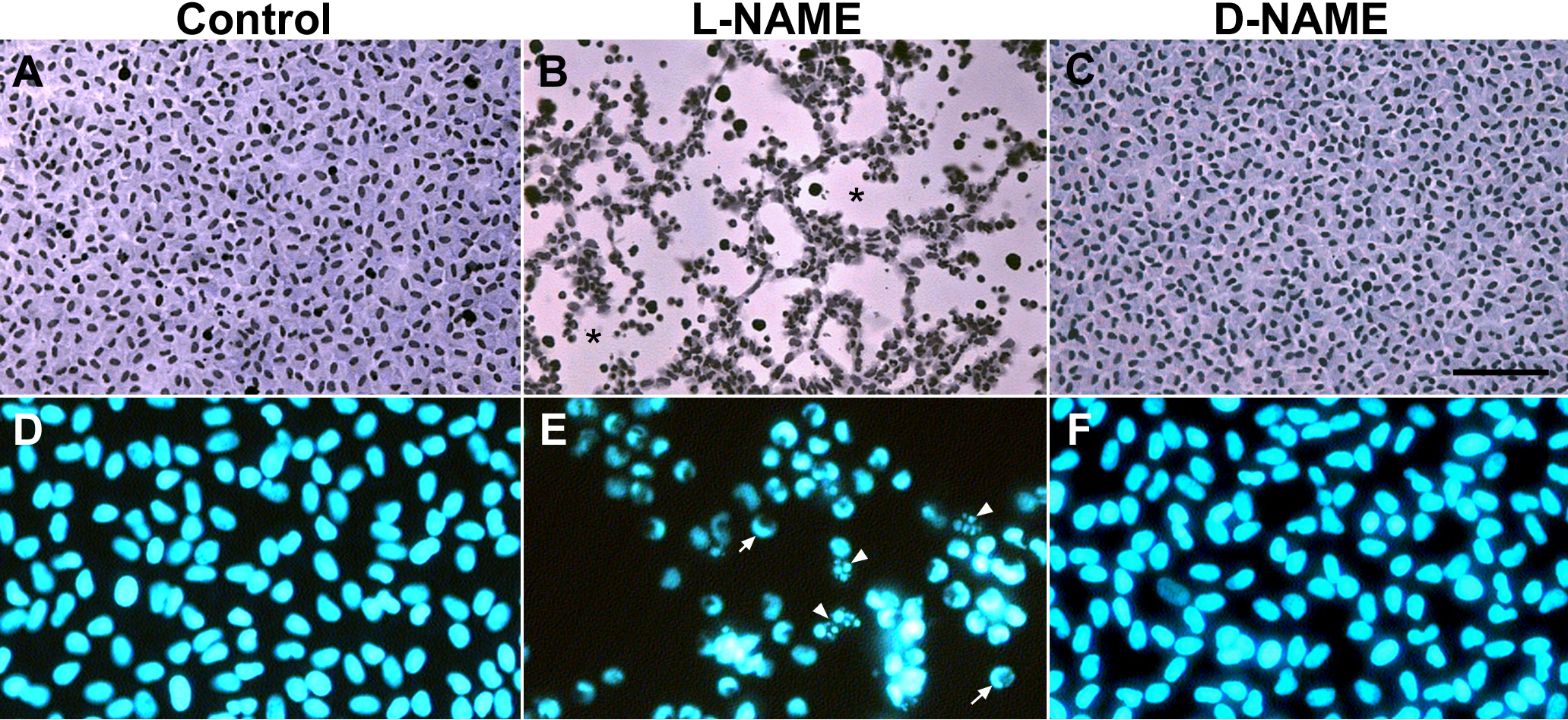
Figure 2. Histology of lens epithelial
explants cultured with the nitric oxide inhibitor L-NAME. Explants were
cultured in the control medium (A and D) or in the
control medium supplemented with 5 mM L-NAME (B and E)
or D-NAME (C and F). Representative explants were fixed
on day 5 of culture and stained with methylene blue-hematoxylin (A-C)
and Hoechst dye (D-F). Explants cultured in the control
medium (A) or the control medium with D-NAME (C) were
completely covered with a monolayer of closely packed cells in the
cobblestone array typical of the normal lens epithelium whereas in the
L-NAME-treated explants (B) large regions of lens capsule were
denuded of cells (denoted by an asterisk). Nuclei in explants cultured
in the control medium alone and the D-NAME-treated explants were
relatively uniform in shape and evenly stained with Hoechst dye (D
and F) whereas nuclei in the L-NAME-treated explants (E)
showed crescent-like staining with Hoechst dye (arrow) or nuclear
fragmentation (arrowheads). The bar represents 80 μm in A-C
and 35 μm in D-F. NAME stands for Nω-nitro-L-arginine
methyl ester.