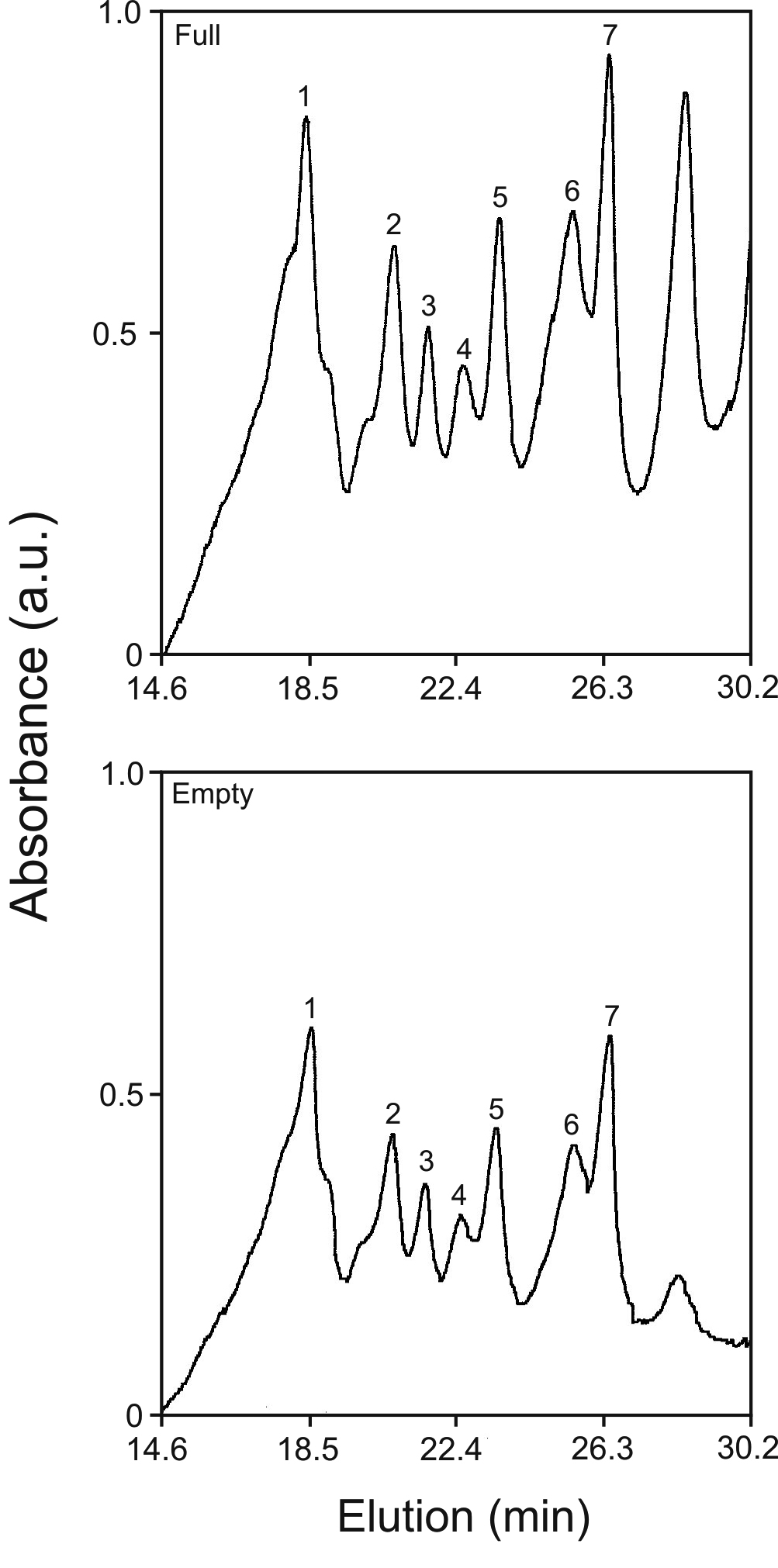
Figure 2. Fetal γ-crystallin binding to
old α-crystallins. Elution profiles of γ-crystallins in the full (top)
versus empty (bottom) chambers of microequilibrium dialysis of old
α-crystallins and fetal calf γ-crystallins. See Methods for details of C
18
reverse phase chromatography to resolve γ-crystallin peaks. Numbered
peaks were quantitated and used to compute binding ratios listed in
Table 1.