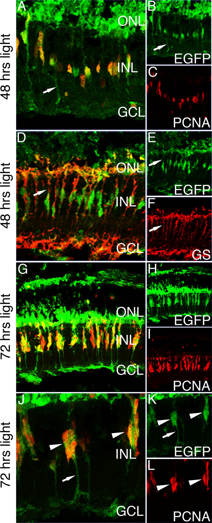
Figure 5. Enhanced green fluorescent
protein expression in the light-damaged Tg(ccnb1:EGFP)nt18
zebrafish retina. To initiate the regeneration response, dark-adapted
adult Tg(ccnb1:EGFP)nt18 zebrafish were
exposed to constant, high-intensity light to induce photoreceptor
apoptosis. At 48 h of constant light exposure, enhanced green
fluorescent protein (EGFP) is expressed in proliferating cell nuclear
antigen (PCNA)-positive inner nuclear layer (INL) cells (A-C).
EGFP expression is also observed in Müller glial cell processes
(arrows). At 48 h of light damage, Müller glia are labeled with
glutamine synthetase (GS; D, F). All the EGFP-positive cells
co-label with glutamine synthetase expressing Müller glia (D-F).
At 72 h of light damage, there are clusters of PCNA-positive neuronal
progenitor cells associated with and migrating along Müller glia (G-I).
These cells express EGFP at a high intensity. (J-L) A higher
magnification of the migrating progenitor cells at 72 h of light damage
reveals that EGFP is expressed throughout the glial cell (J, K).
This is evident by labeling in the glial cell processes (arrows). There
are multiple PCNA-positive progenitors that are also expressing EGFP
(arrowheads). Abbreviations: GS represents glutamine synthetase; PCNA
represents proliferating cell nuclear antigen; EGFP represents enhanced
green fluorescent protein; ONL represents outer nuclear layer; INL
represents inner nuclear layer; and GCL represents ganglion cell layer.