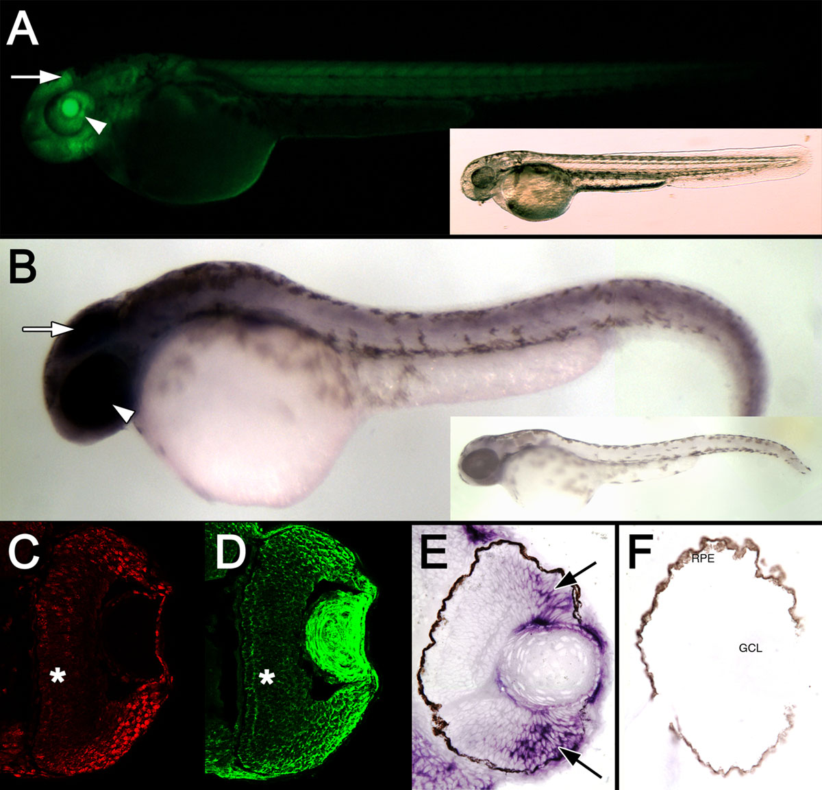
Figure 2. Expression of the cyclin
B1:EGFP transgene at 48 hpf. A: A fluorescent image of a Tg(ccnb1:EGFP)nt18
embryo at 48 hpf is displayed. EGFP was expressed throughout the body
of the fish with the most intense expression in the brain (arrow) and
eye field (arrowhead). A bright-field image of the same embryo is shown
in the inset. B: Whole-mount in situ hybridization revealed cyclin
B1 mRNA expression throughout the body with the greatest staining
in the head (arrow) and eye (arrowhead). A zebrafish labeled with the cyclin
B1 sense RNA probe is shown in the inset. Retinal sections at 48
hpf reveal strong PCNA expression in cells in the retinal margin (C),
which is similar to EGFP expression (D). EGFP and PCNA
expression is decreased in the central retina (*). Retinal sections
labeled with the cyclin B1 antisense RNA probe demonstrated a
similar high level of expression near the retinal margin (E,
arrows), while the control sense RNA probe did not label any retinal
cells. F: No signal was observed with the sense RNA probe.
Abbreviations: RPE represents retinal pigmented epithelium, and GCL
represents ganglion cell layer.