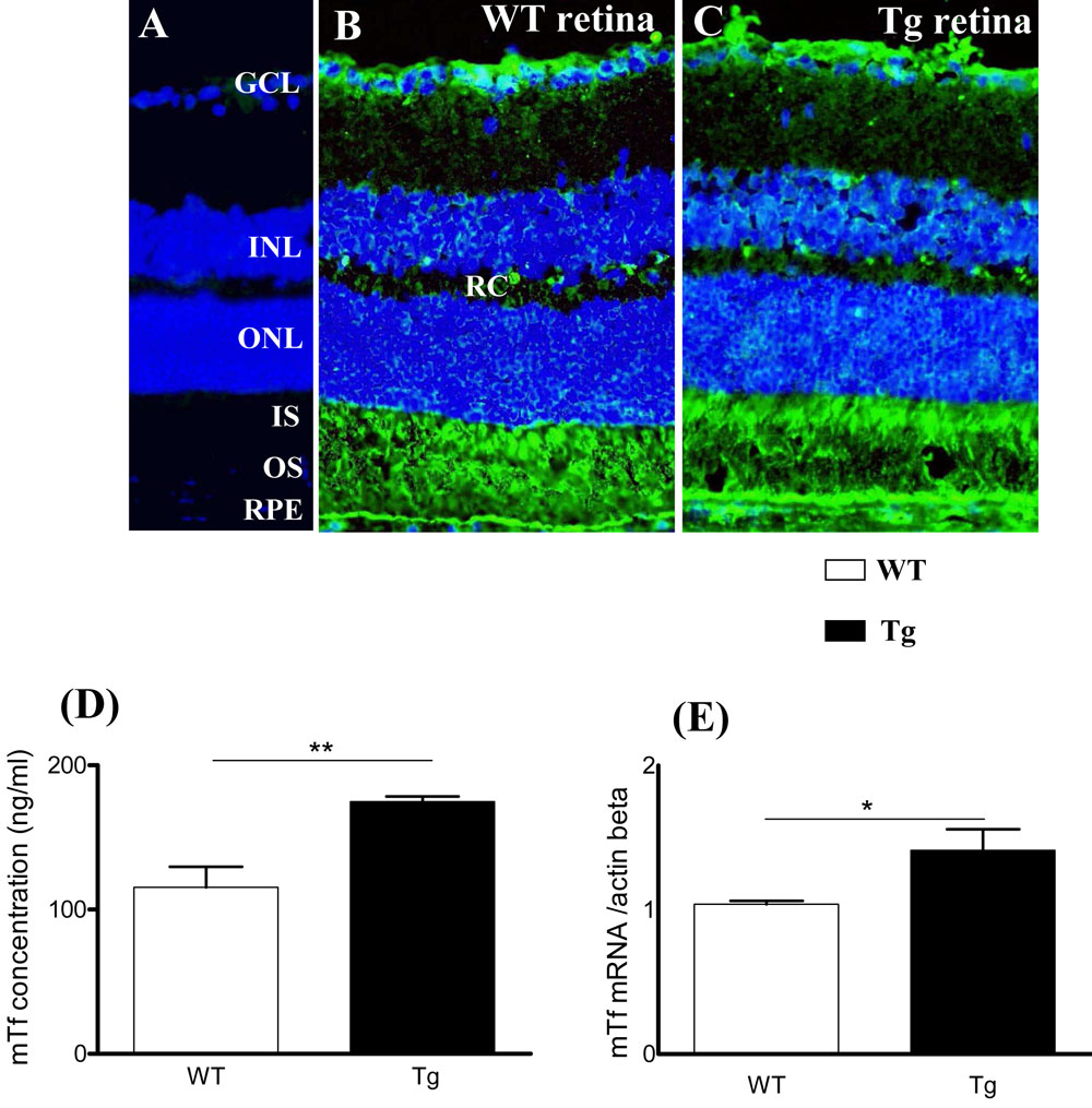
Figure 2. Mouse transferrin is synthetized
and localized in one-month-old mouse retina. A: Non-immune serum was
used as a control. B-C: Mouse transferrin (mTf; green) is localized on
frozen sections of wild-type (WT) mice (A,B) and in transgenic
(Tg) mice (C). Scale bar equals 100 µM. The following
abbreviations are used: Ganglion cell layer (GCL), inner nuclear layer
(INL), inner plexifrom layer (IPL), inner segments (IS), outer limiting
membrane (OLM), outer nuclear layer (ONL), outer plexifrom layer
(OPL), outer segments (OS), retinal capillaries (RC), retinal pigmented
epithelium (RPE). D: mTf concentration (ng/ml) was measured by
radioimmunoassay (RIA) in supernatant of retinal extracts of WT and Tg
mice . Each column represents the mean ±SEM. The double asterisk
represents statistical significance of differences from control,
p<0.01. E: mTf mRNA expression was quantified by RT-qPCR in retinal
extracts from WT and Tg mice. Each column represents the mean ±SEM. The
asterisk represents statistical significance of differences from
control, p<0.05.