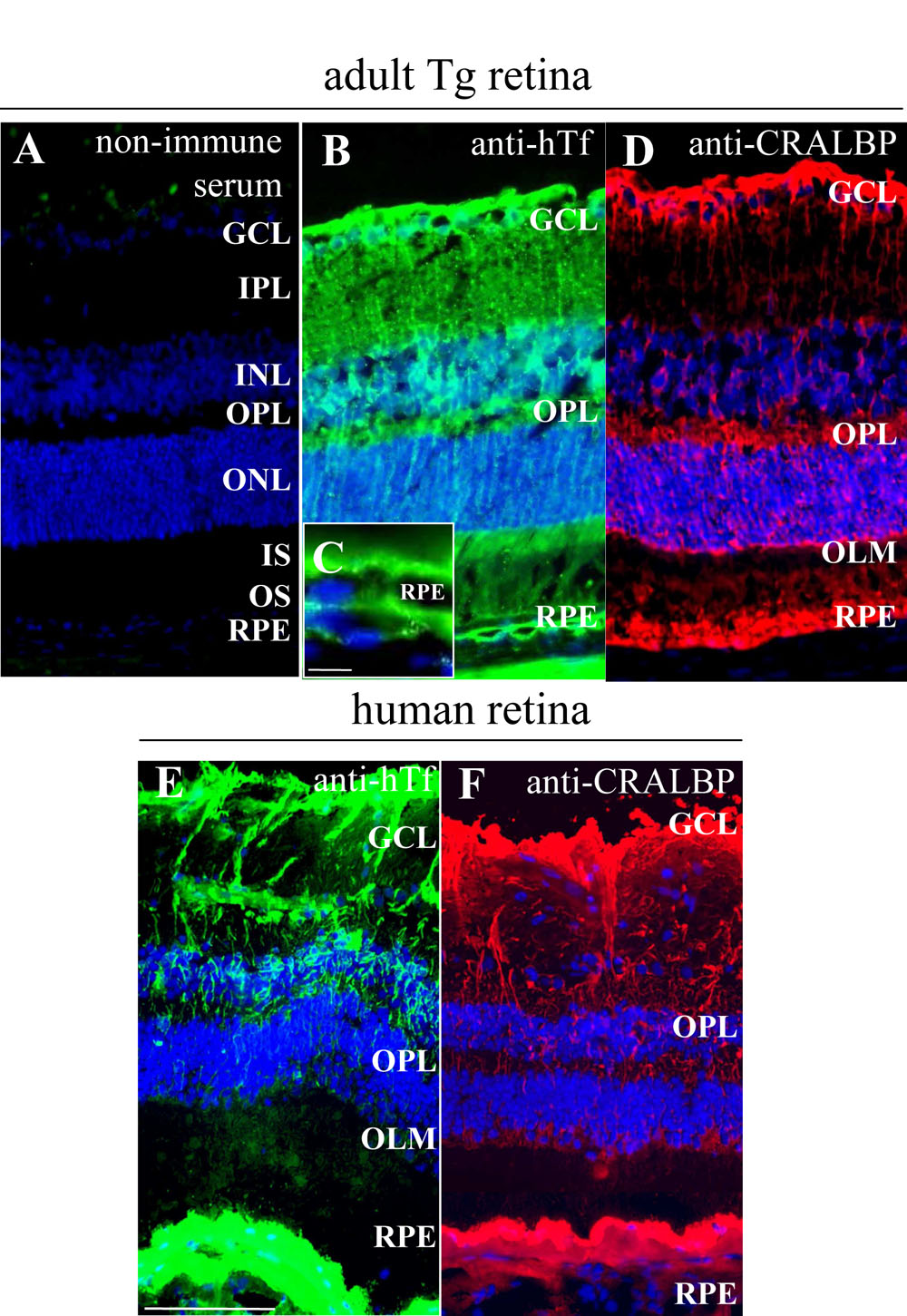
Figure 1. Human transferrin is localized
in the same cells in transgenic mice retina as in human retina. A:
Non-immune serum was used as a control. B-D: Human transferrin (hTf;
green) localization was realized on frozen sections of one-month-old
transgenic (Tg) mice retina. hTf is localized in Müller glial (MG)
cells and retinal pigment epithelium (RPE; B,C), identified
with cellular retinaldehyde binding protein (CRALBP; red; D),
specific marker. C: Higher magnification of RPE from (B). E-F:
hTf is localized in a 90-year-old human retina (E) in the same
cells as in Tg mice still identified by CRALBP (F). The
following abbreviations are used: Ganglion cell layer (GCL), inner
nuclear layer (INL), inner plexifrom layer (IPL), inner segments
(IS), outer limiting membrane (OLM), outer nuclear layer (ONL),
outer plexifrom layer (OPL), outer segments (OS), retinal pigmented
epithelium (RPE). The scale bars in A,B, D-F
equal 100 µM and the scale bar in C equals 10 µm.