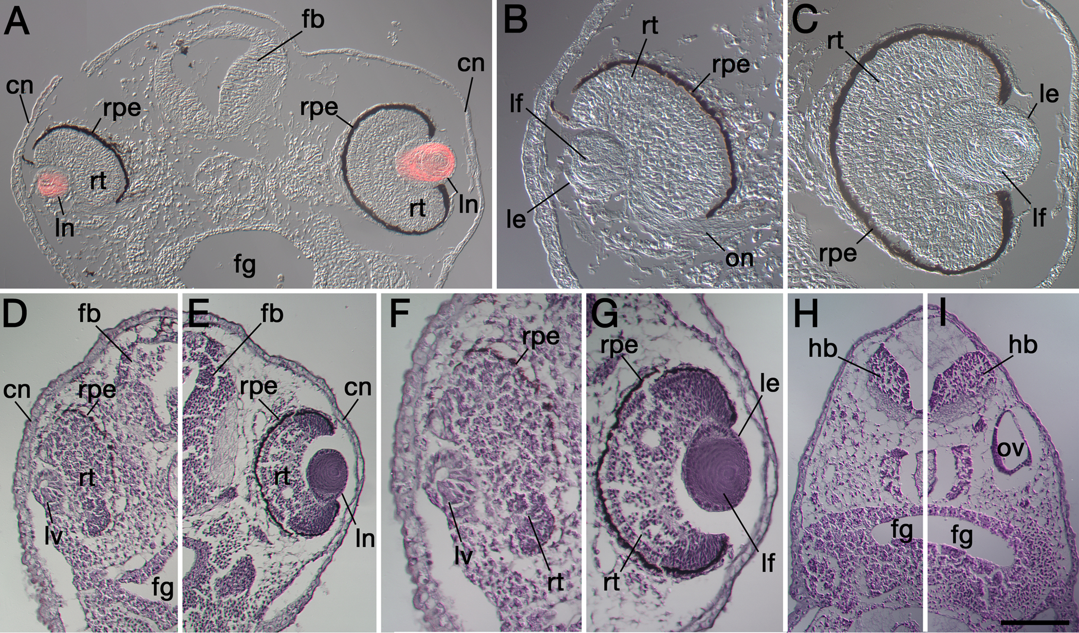
Figure 4. Transverse sections of specimens
demonstrating severe eye defects following Psf2MO-mediated knockdown.
Dorsal is toward the top in each figure. A: Image montage of a
typical specimen shows the Psf2 morpholino-injected side on the left
and the internal control, uninjected side on the right. This image was
viewed with differential interference contrast (DIC). The red color in A
shows overlain distribution of rhodamine fluorescence of secondary
antibodies revealing immunoreactive lens crystallin proteins within
both lenses. The cornea epithelium overlying the eye on the left is
thicker compared to the uninjected side on the right, characteristic of
undifferentiated embryonic ectoderm. Note that the lens and retina of
the morpholino-affected eye are smaller and not as fully
differentiated. The forebrain is also smaller and less differentiated
on the left, Psf2MO-injected side. B and C are the
higher magnification views of the left and right eyes shown in A,
respectively. D-E: The left and right sides of the head
of a second case stained with Hematoxylin/Eosin are shown. F
and G are higher magnification views of the eyes shown in D
and E, respectively. Note that the forebrain and retina are
malformed on the left, Psf2MO-injected side shown in D and F
compared to the control side shown in E and G. On the
Psf2MO-injected side (D, F), only a small lens vesicle
possessing a central lumen has formed. This lens vesicle exhibits some
polarization and evidence of elongating primary fiber cells. Also note
the retarded differentiation of the cornea in D and F
compared to the respective control cornea shown in E and G.
H-I: More posterior sections show the reduction in
hindbrain size and the absence of the otic vesicle on the
Psf2MO-injected side (H) compared to the normal pattern of
development seen on the control, uninjected side (I). cn, cornea
epithelium; fb, forebrain; fg, foregut; hb, hindbrain; le, lens
epithelium; lf, lens fiber cells; ln, lens; lv, lens vesicle; on, optic
nerve; ov, otic vesicle; rpe, retinal pigmented epithelium; rt, neural
retina. The scale bar in I is equal to 160 µm in A-B,
85 µm in B-C, 170 µm in D-E, 100 µm in F-G,
and 190 µm in H-I.