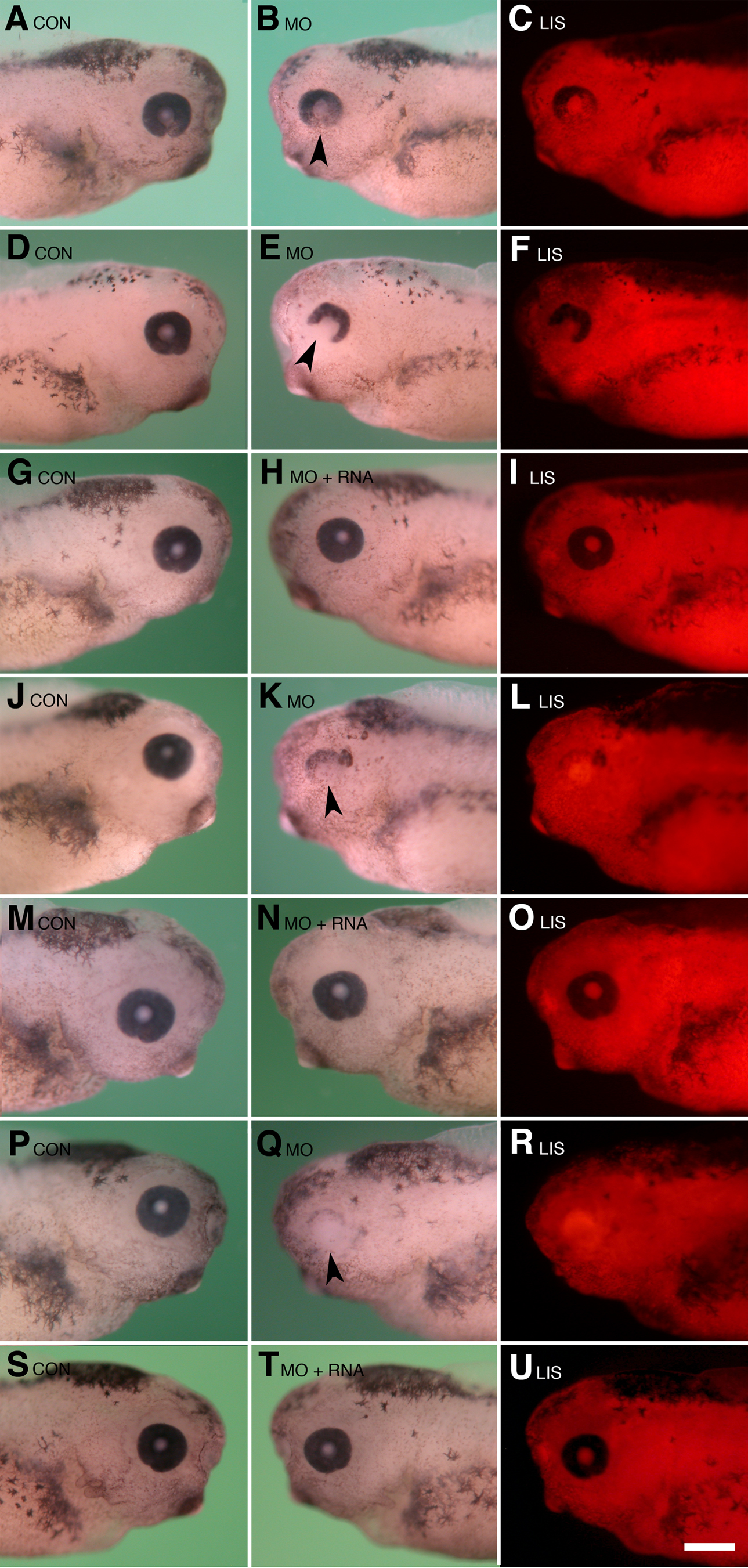
Figure 3. Results of Psf2
morpholino knockdown and RNA rescue experiments. Dorsal is toward the
top in each figure. A-F: Typical eye defects observed
following unilateral injection of lissamine-tagged Psf2MO into single
blastomeres at the two-cell stage (equivalent to 2.25 ng/cell at the
eight-cell stage) are shown. A: Normal control (uninjected,
CON) side is shown. This side is the normal part the embryo shown in B-C.
B: Minor eye defect is observed on the Psf2MO-injected side
(opposite that shown in A) as indicated by arrow. Note the
slight decrease in retinal pigmentation in the ventral region and the
reduced size of the optic cup compared to that shown in A. C:
Corresponding fluorescence image to that shown in B, the image
shows the distribution of the lissamine-tagged morpholino (LIS). D:
Normal control, uninjected side is shown. This side is the normal part
of the embryo shown in E-F. E: A severe eye
defect phenotype is observed following Psf2MO injection. Note the
smaller size of the eye and severe reduction in the ventral region of
the optic cup, denoted by the arrow in E. F: The
corresponding fluorescence image shows the distribution of the
lissamine-tagged morpholino for the embryo shown in E. G-I:
The typical result observed following the co-injection of 2.25 ng Psf2
morpholino and 1000 pg synthetic altPsf2 RNA is shown. G: The
normal control, uninjected side of the embryo shown in H-I
is displayed. H: There was normal morphological development
following the co-injection of Psf2MO (equivalent to 2.25 ng/blastomere
at the eight-cell stage) and 1000 pg rescue RNA (altPsf2 RNA). I:
The corresponding fluorescence image to that shown in H shows
distribution of the lissamine-tagged morpholino. J-L:
Typical severe eye defect is observed following the unilateral
injection of lissamine-tagged Psf2MO into single blastomeres at the
two-cell and four-cell stages (equivalent to 4.5 ng/blastomere at the
eight-cell stage). J: The normal control, uninjected side of
the embryo shown in K-L is pictured. K: Typical
severe eye defect phenotype is observed following Psf2MO injection.
Note the smaller size of the eye and severe reduction in the ventral
region of the optic cup, denoted by the arrow in K. L:
The corresponding fluorescence image shows the distribution of the
lissamine-tagged morpholino for the embryo shown in K. M-O:
The typical result is observed following the co-injection of 4.5 ng Psf2
morpholino and 1000 pg synthetic altPsf2 RNA. M: The normal
control, uninjected side of the embryo shown in N-O is
displayed. N: Normal morphological development is observed
following the co-injection of Psf2MO (equivalent to 4.5 ng/blastomere
at the eight-cell stage) and 1000 pg rescue RNA (altPsf2 RNA). O:
The corresponding fluorescence image to that shown in N shows
the distribution of the lissamine-tagged morpholino. P-R:
Typical severe eye defect is observed following unilateral injection of
lissamine-tagged Psf2MO into single blastomeres at the two-cell and
four-cell stages (equivalent to 6 ng/blastomere at the eight-cell
stage). P: The normal control, uninjected side of the embryo
shown in Q and R is shown. Q: The typical
severe eye defect phenotype is displayed following Psf2MO injection.
Note the severe reduction in the ventral region of the optic cup,
denoted by the arrow in Q, and the overall lack of retinal
pigmentation. R: The corresponding fluorescence image shows the
distribution of the lissamine-tagged morpholino for the embryo shown in
Q. S-U: The typical result is observed following
co-injection of 6 ng Psf2 morpholino and 1000 pg synthetic
altPsf2 RNA. S: The normal control, uninjected side of the
embryo shown in T-U is displayed. T: There is
normal morphological development following the co-injection of Psf2MO
(equivalent to 6 ng/blastomere at the eight-cell stage) and 1000 pg
rescue RNA (altPsf2 RNA). U: The corresponding fluorescence
image to that shown in S shows the distribution of the
lissamine-tagged morpholino. The scale bar in U is equal to 450
µm.