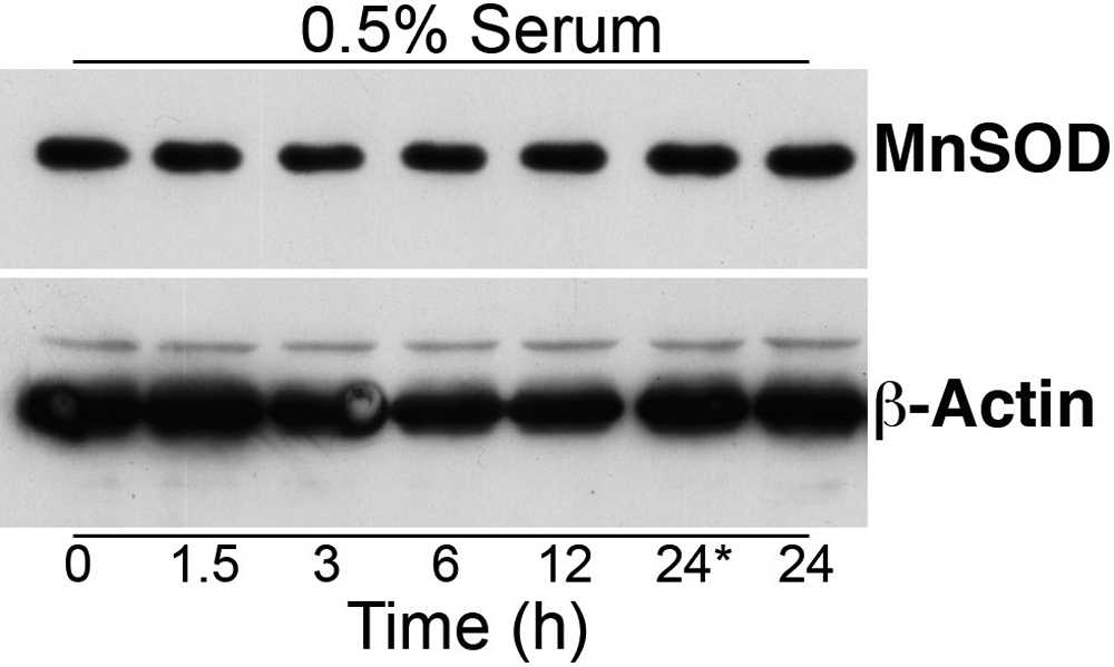
Figure 3. Western blot analysis of
manganese superoxide dismutase expression in HLE-B3 cells. Total cell
lysates were collected from HLE-B3 cells grown in 0.5% serum and
stimulated by exposure to 1 μM 17β-E2 for 0, 1.5, 3, 6, 12,
and 24 h. There was no change in the expression of MnSOD at any of the
time points. The time points at 0 and 24* hours did not receive
estrogen.