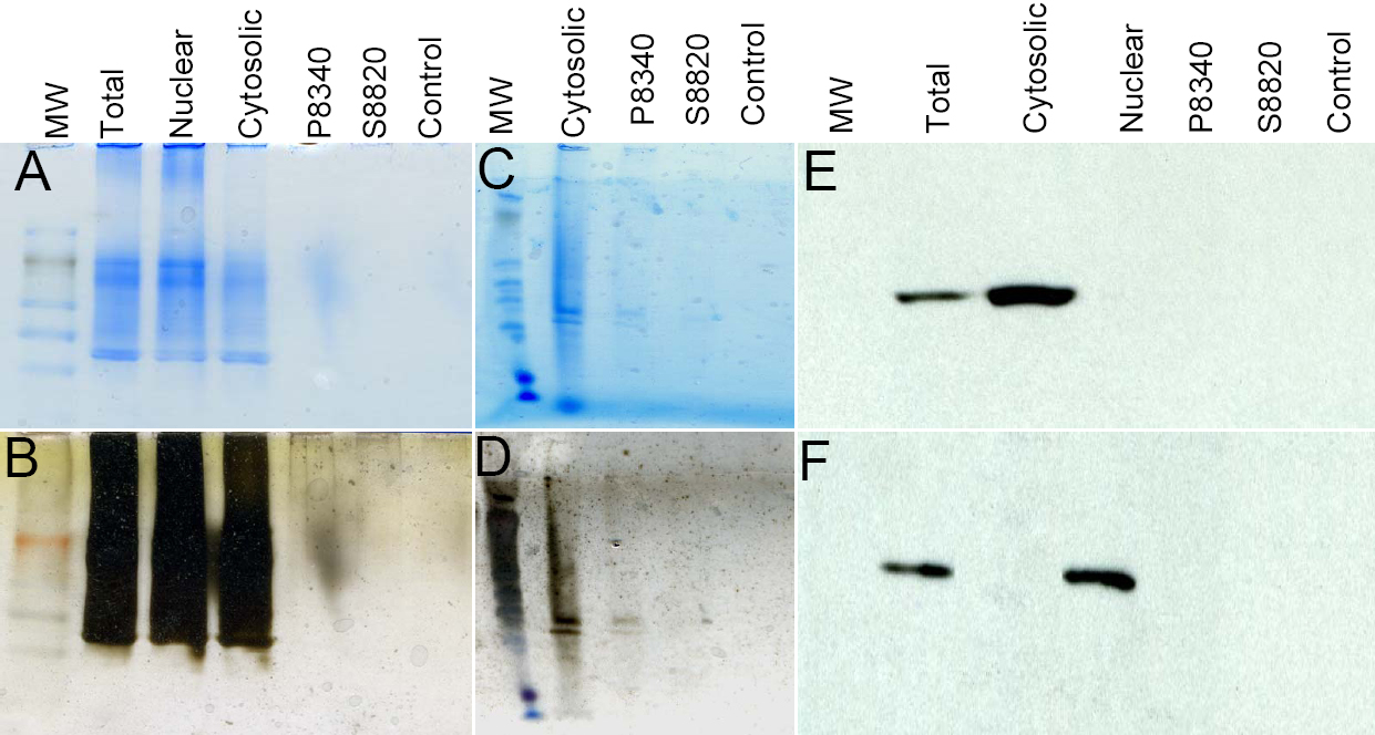
Figure 1. Analyses of SDS–PAGE
fractionated proteins in enrichment steps. A. About 100 µg of
protein (initial load) was subjected to affinity enrichment on P8340 or
S8820 column as indicated and total eluted and acetone precipitated
proteins were subjected to SDS–PAGE fractionation and stained with
Coomassie blue. An initial load (10 µg) of total, nuclear and cytosolic
proteins were fractionated on a SDS–PAGE. For this purpose cytosolic
and nuclear fractions (10 µg proteins each) from NE-PER Nuclear and
Cytoplasmic Extraction Reagents kit (Pierce Biotechnology Inc.) were
obtained B. The same gel as in A was subjected to silver
staining. C. About 250 µg of protein (initial load) was
subjected to affinity enrichment on P8340 or S8820 column as indicated.
Total eluted and acetone precipitated proteins were subjected to
fractionation on a 4%–15% PHAST gel (GE Healthcare) and stained with
Coomassie blue. D. The same gel as in C was subjected to silver
staining. The control is the cytosolic fraction (4 µg) passed through
an empty protein A column as described in methods. E. After
transfer to a PVDF membrane, western analyses of protein extracts were
performed, using GAPDH antibody and, F. Histone H3 antibody, as
described in methods.