![]() Figure 4 of
Papanikolopoulou, Mol Vis 2008;
14:81-89.
Figure 4 of
Papanikolopoulou, Mol Vis 2008;
14:81-89.
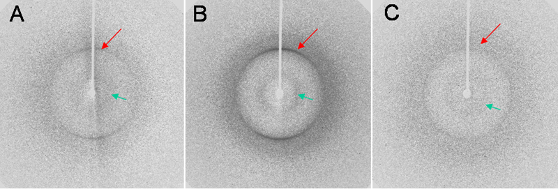
Figure 4. X-ray fiber diffraction from crystallin fibrils
X-ray fiber diffraction patterns recorded for the A: HγD-Crys, B: HγD-Crys Ctd, and C: HγD-Crys Ntd fibrils showing the characteristic features associated with the cross-β amyloid motif: an H-bonding 4.7 Å (long arrow) meridional reflection and an about 10 Å broad reflection (short arrow) on the equator. See experimental procedures for sample preparation.