![]() Figure 2 of
Papanikolopoulou, Mol Vis 2008;
14:81-89.
Figure 2 of
Papanikolopoulou, Mol Vis 2008;
14:81-89.
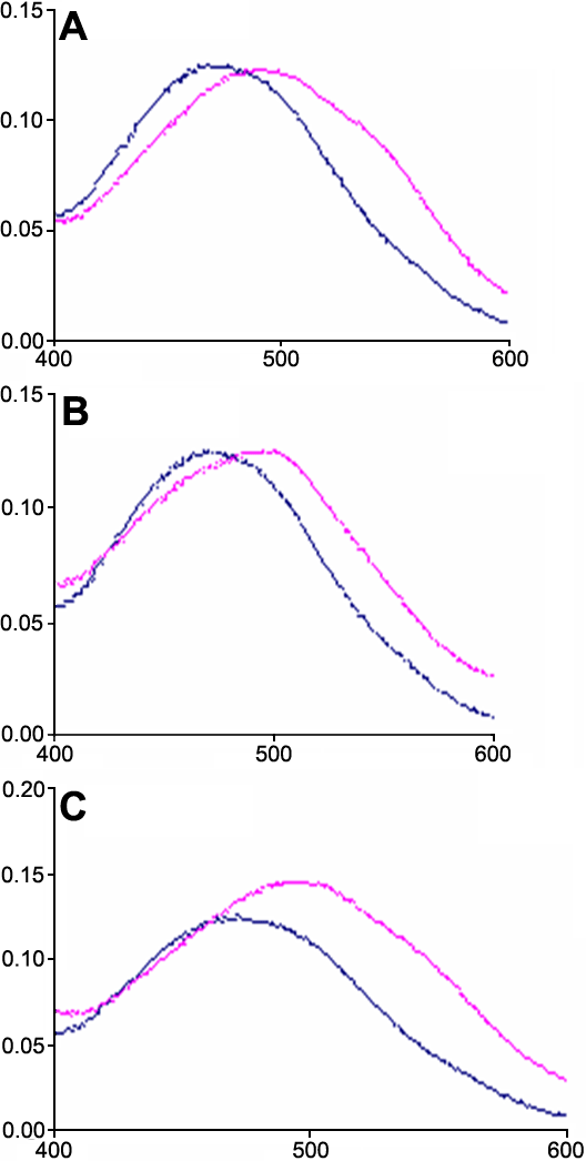
Figure 2. Congo red binding to crystallin fibrils
The spectra of 5 μM Congo red in the absence (blue line) and in the presence (red line) of fibrils formed by A: HγD-Crys, B: HγD-Crys Ctd, and C: HγD-Crys Ntd. Before adding the dye, the scattering of the peptide solutions was recorded and subtracted from the spectrum of the dye in their presence. Fibrils were formed by incubation of the proteins at 37 °C in 50 mM acetate buffer pH 3 at 5 mg/ml for 2 days and subsequently diluted in a 500 mM sodium phosphate buffer pH 7 at a 0.5 mg/ml concentration.