![]() Figure 1 of
Papanikolopoulou, Mol Vis 2008;
14:81-89.
Figure 1 of
Papanikolopoulou, Mol Vis 2008;
14:81-89.
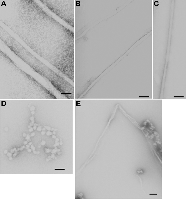
Figure 1. Electron micrographs of fibrils negatively stained with 1% uranyl acetate
Conditions were as follows: A: 5 mg/ml solution of the HγD-Crys into 50 mM acetate buffer pH 3 incubated at 37 °C for 2 days, B: 5 mg/ml solution of the HγD-Crys Ctd into 50 mM acetate buffer pH 3 incubated at 37 °C for 2 days, C: 5 mg/ml solution of the HγD-Crys Ntd into 50 mM acetate buffer pH 3 incubated at 37 °C for 2 days, D: 50 μg/ml solution of the HγD-Crys into 100 mM sodium citrate pH3 deposited on the grid after 2 h of incubation at 37 °C, E: 50 μg/ml solution of the HγD-Crys into 100 mM sodium citrate pH3 deposited on the grid after 6 h of incubation at 37 °C. The bar represents 1,000 Å.