![]() Figure 7 of
Hitani, Mol Vis 2008;
14:1-9.
Figure 7 of
Hitani, Mol Vis 2008;
14:1-9.
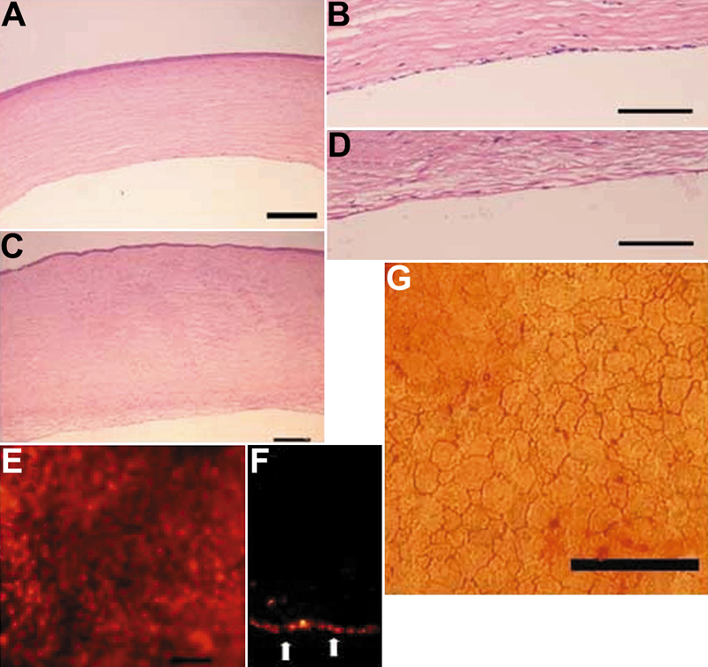
Figure 7. Histologic examination and localization of human corneal endothelial cells after transplantation
A and B: In the HCEC sheet group, a monolayer of continuous cells attached to the posterior surface of a rabbit cornea. C and D: On the other hand, in the control group, there was no Descemet's membrane, and endothelial cells on the posterior surface of rabbit corneas as well as the corneal stroma were thickened. E and F: Fluorescein microscopic examination of whole mount corneas (E) and thin sections (F) showed that the posterior surface of a transplanted cornea was covered with PKH26-labeled cells and apparent cell defects were not observed on the HCEC cell sheet. G: The average cell density in the HCEC sheet group 7 days postoperatively was 2,244 cells/mm2. The scale bars in A, C, and G are equal to 100 μm, 200 μm in B and D, and 50 μm in E.