![]() Figure 4 of
Hitani, Mol Vis 2008;
14:1-9.
Figure 4 of
Hitani, Mol Vis 2008;
14:1-9.
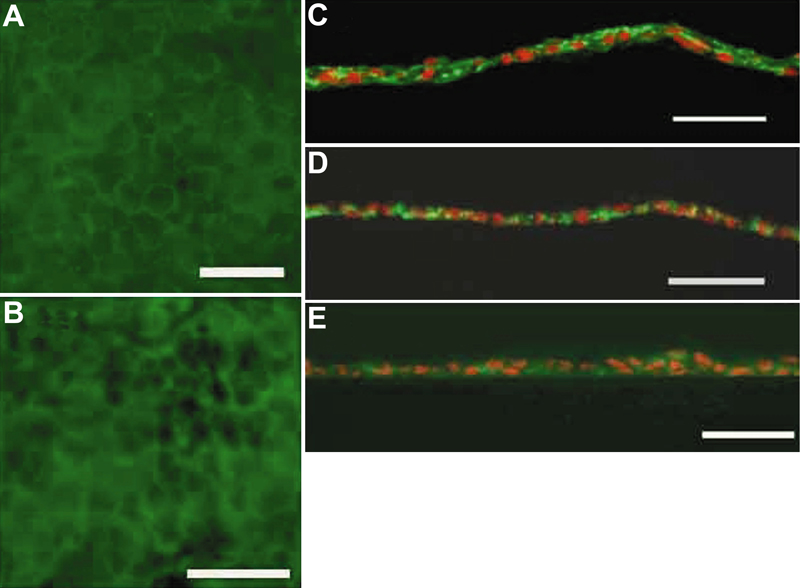
Figure 4. Immunohistologic findings of the human corneal endothelial cell sheet
A and B: In the flat mount immunohistochemical observation, immunostaining of both ZO-1 (A) and Na+, K+-ATPase (B) were positive at most cellular boundaries. The cellular shape demarcated with immunostaining was quasi-regular with well defined cell boundaries. C: In the transverse observation of extracellular matrix, immunostaining for laminin was positive at the basal side, superficial side, and intercellular space. D and E: Immunostaining of fibronectin (D) and type IV collagen (E) was weak at the surface of the sheet, but positive at the intercellular space. The scale bars are equal to 50 μm.