![]() Figure 3 of
Hitani, Mol Vis 2008;
14:1-9.
Figure 3 of
Hitani, Mol Vis 2008;
14:1-9.
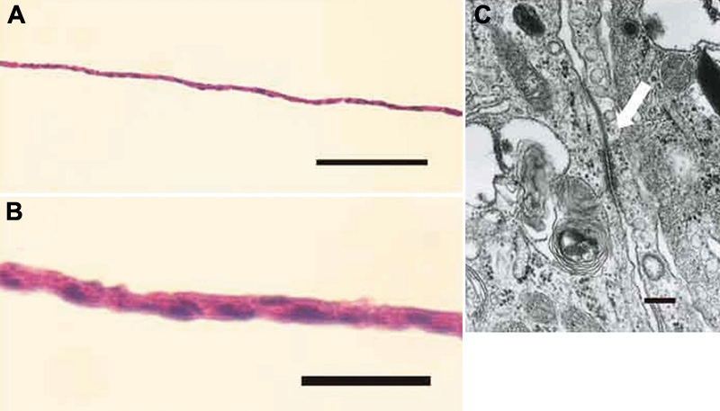
Figure 3. Histologic findings of the human corneal endothelial cell sheet
A and B: A cell sheet consists of a monolayer of cells that have consistent size. The scale bar in A is equal to 200 μm and in B is equal to 50 μm. C: Electron microscopic observation demonstrated desmosomes between cells (arrow). Bar=200 nm.