![]() Figure 2 of
Hitani, Mol Vis 2008;
14:1-9.
Figure 2 of
Hitani, Mol Vis 2008;
14:1-9.
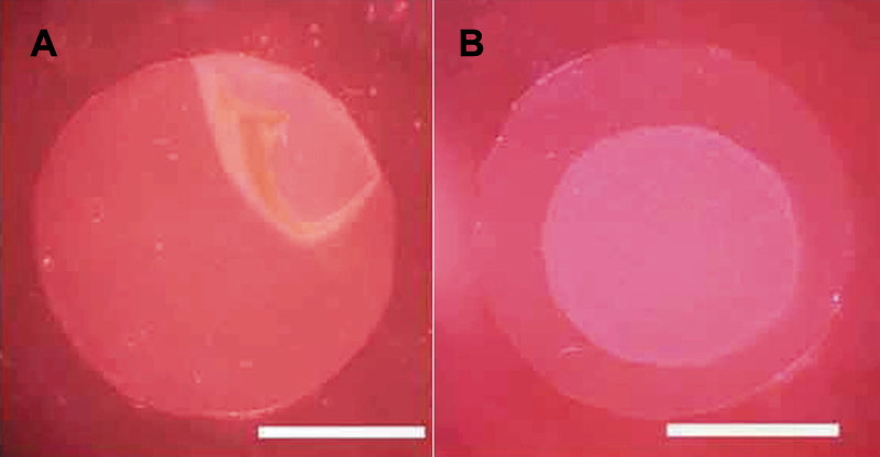
Figure 2. Preparation of a human corneal endothelial cell sheet
A: Cultured HCECs could be bluntly detached en bloc from the bottom of a culture insert using a spatula after EDTA treatment to the bottom side of the culture insert. B: The detached sheets had a circular shape with an approximately 6 mm diameter. The scale bars are equal to 5 mm.