![]() Figure 9 of
Jakobs, Mol Vis 2007;
13:933-948.
Figure 9 of
Jakobs, Mol Vis 2007;
13:933-948.
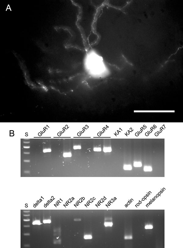
Figure 9. Melanopsin-containing ganglion cell
A: The picture shows a projection of 10 image planes taken at 1 mm step size to reveal the dendritic morphology of this melanopsin positive ganglion cell. (Note that this cell was GFP-positive in a GFP-M retina. Therefore the morphology is obvious without immunostaining.) the scale bar represents 50 μm. B: Agarose gels showing the iGluR expression in this one cell, which was positive for GluR1flop, GluR2flop, GluR3flip, GluR4flip and flop, KA2, GluR5, GluR6, δ1 and δ2, NR1, NR2c, NR3a. In addition, this cell was assayed for the expression of melanopsin.