![]() Figure 5 of
Jakobs, Mol Vis 2007;
13:933-948.
Figure 5 of
Jakobs, Mol Vis 2007;
13:933-948.
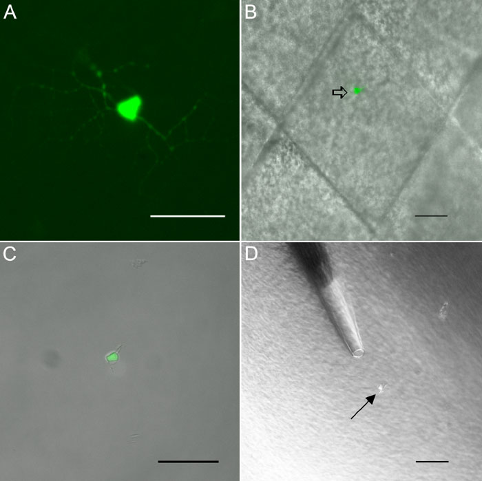
Figure 5. Microdissection procedure for morphologically characterized ganglion cells
A: GFP-labeled ganglion cell. Before isolation ganglion cells from GFP-M retinas were photographed and their level of stratification in the IPL was measured. B: a piece of tissue (about 100 mmx100 mm) containing only this one labeled ganglion cell was cut out. DIC and GFP images of the same piece are superimposed. Double arrow indicates the targeted ganglion cell. C: After dissociation the fluorescent ganglion cell was picked up from the cell suspension and transferred to another well with fresh buffer for washing. DIC and GFP pictures for the same isolated cell are superimposed. D: Finally the cell was transferred directly into the reaction tube containing PCR buffer. The transfer pipette is visible in the upper half of the picture, the arrow indicates the cell. All scale bars equal 50 μm.