![]() Figure 7 of
Johnson, Mol Vis 2007;
13:887-919.
Figure 7 of
Johnson, Mol Vis 2007;
13:887-919.
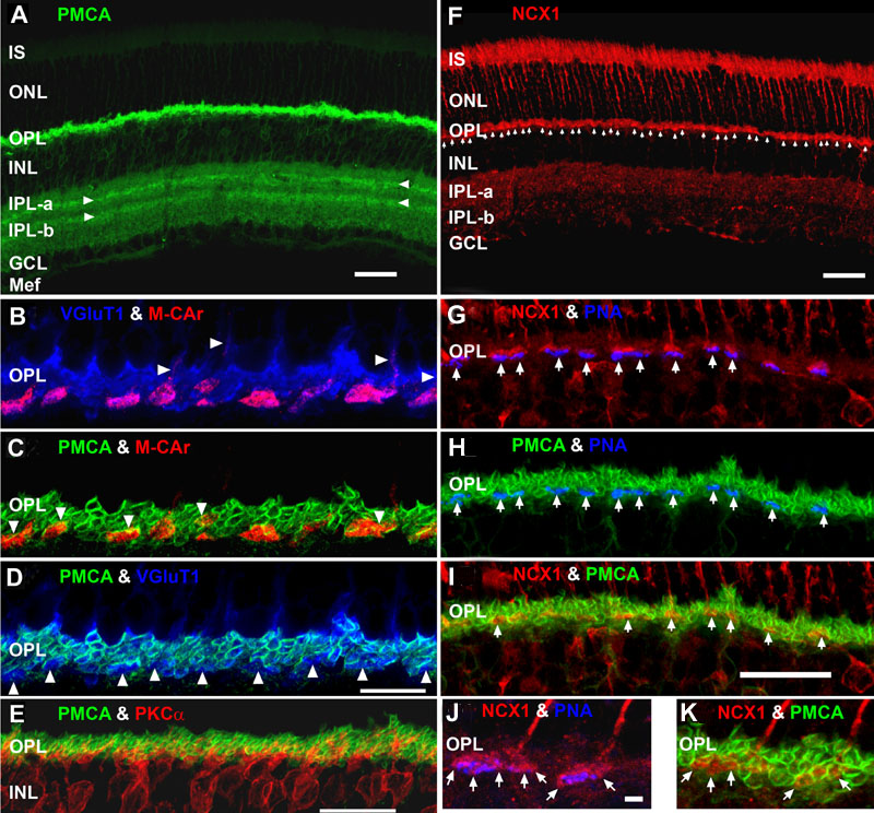
Figure 7. Lamination patterns and differential compartmentation of pan-plasma membrane Ca2+ ATPase and Na+-Ca2+ exchanger isoform 1 in rod spherules and cone pedicles
A: Confocal image of retina immunolabeled for pan-plasma membrane Ca2+ ATPase (pan-PMCA). The double white arrowheads identify strongly labeled PMCA bands in each IPL sublamina. The scale bar applies to A, D and represents 40 μm. B-D: PMCA preferentially labels rod spherules. OPL double labeled with markers for PMCA (green), vesicular glutamate transporter 1 (VGluT1: blue) and/or M-cone arrestin (M-CAr: red). Scale bar equal 20 μm. B: VGluT1, which labels photoreceptor terminals and M-CAr, which labels cones, colocalize in the OPL. This reveals the large dome-shaped cone pedicles (purple pixels) and some cone axons (white arrowheads). C: This high magnification image shows the horseshoe-like appearance of the PMCA-positive rod spherules. In contrast, the retina double labeled with PMCA and M-CAr shows that cone pedicles stain weakly and diffusely for PMCA, except for a discrete band at the top of the pedicle (white arrowheads: yellow pixels). D: A retina double labeled with PMCA and VGluT1 confirms that PMCA extensively labels rod spherules, but only sparsely labels cone pedicles (white arrowheads). E: PKCα-positive rod bipolar cells (red) are pan-PMCA-negative (green), although they are in close apposition around the rod spherules (yellow pixels). The scale bar represents 20 μm. F: Retinal localization of Na+-Ca2+ exchanger isoform 1 (NCX1). NCX1 intensely labels IS, cone axons and cone pedicles (white arrowheads). G-I: NCX1 preferentially labels cone pedicles. OPL from triple labeled experiments with markers for NCX1 (red), PMCA (green), and/or PNA (blue). Scale bar equal 20 μm. G: PNA stains selected flat contact regions on the large cone terminal (white arrows). NCX1 intensely labeled the entire cone pedicle and axon terminals, whereas rod spherules were diffusely labeled. H: PMCA labels the rod spherule membranes with a horseshoe-like appearance, but does not prominently colocalize with PNA-positive cone pedicle membranes (white arrows). I: Retina double labeled with PMCA and NCX1 shows that these proteins colocalized in cone pedicles near the axon junction (white arrows: yellow pixels). J, K: High magnification confocal images showing preferential labeling of cone pedicles by NCX1 and rod spherules by PMCA. OPL from triple labeled experiments with markers for NCX1 (red), PMCA (green), and/or PNA (blue). Scale bar equals to 10 μm. J: Double labeled retina shows that NCX1 intensely labels the entire cone pedicle and axon terminals, but only diffusely labels the rod spherules. This high magnification image also shows that PNA stains selected flat contact regions on the large cone terminal, whereas invaginating synaptic regions (white arrows) are not labeled by PNA. K: Double labeled retina shows that PMCA and NCX1 colocalize in cone pedicles near the axon junction (white arrows: yellow pixels).