![]() Figure 6 of
Johnson, Mol Vis 2007;
13:887-919.
Figure 6 of
Johnson, Mol Vis 2007;
13:887-919.
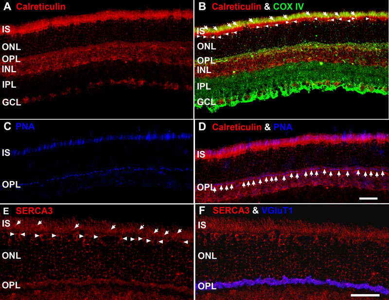
Figure 6. Molecular markers of endoplasmic reticulum (ER) reveal distinct retinal distribution and lamination patterns
A-D: Confocal image of retina triple labeled for calreticulin, COX IV and PNA. Scale bar equal 20 μm. A: Retina stained for calreticulin, a Ca2+-binding protein present in all ER lumen. Note the intense labeling in the IS, OPL and INL. B: Location of calreticulin (red) and COX IV (green). These proteins are in close apposition in the ellipsoid region of photoreceptor IS (white arrows and yellow pixels), juxtanuclear mitochondria associated with cone somas (white arrowheads) and in the OPL (punctate yellow-orange pixels). C: Retina stained with peanut agglutinin (PNA), a relatively selective marker for cones, shows distinct labeling in the IS and OPL. D: Calreticulin (red) and PNA (blue) colocalize in CIS and near the pedicles (white arrows; bright purple pixels). E: Pan-Smooth ER Ca2+ ATPase isoform 3 (SERCA3) immunolabeling. This antibody labels in close apposition to the RIS and CIS (white arrows), juxtanuclear mitochondria associated with cone somas (white arrowheads) and the mitochondria in the OPL. F: Colocalization of pan-SERCA3 (red) and VGluT1 (blue) throughout the OPL, indicating the presence of ER in the photoreceptor synaptic terminals. Scale bar for panels E and F equal 40 μm.