![]() Figure 4 of
Johnson, Mol Vis 2007;
13:887-919.
Figure 4 of
Johnson, Mol Vis 2007;
13:887-919.
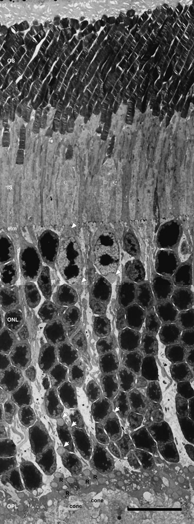
Figure 4. Low-magnification electron micrograph of longitudinal section of the entire photoreceptor layer
Rod and cone (C) photoreceptors are distinguished by several morphological differences. Rod inner segments (IS) are longer, thicker and located more distally in the retina, whereas cone IS are larger and more electron lucent. The cone nuclei are located in the outer third of the outer nuclear layer (ONL), contain several clumps of irregularly shaped heterochromatin and possess two juxtanuclear mitochondria: one above and one below the nucleus (black outlined white arrowheads). The large mitochondrion at the base of the cone nuclei is located at the origin of the wide cone axon, as previously noted [53]. Rod nuclei are present throughout the ONL and contain a single compact mass of heterochromatin. Single juxtanuclear mitochondria are present in many rod somas located in the inner third of the ONL (black outlined white arrows). Numerous synaptic terminals, with large mitochondria, are in the outer plexiform (OPL). Three to five tiers of dark-staining rod spherules (R) overly a single row of more electron lucent cone pedicles (cone) that contain multiple mitochondria. Müller glial cell processes extend throughout the OPL and ONL, and terminate at the external limiting membrane (elm). Scale bar equal 10 μm.